
by MyH Editorial Team | Oct 9, 2023 | Lab Protocols, White Blood Cells
Procedure-At-A-Glance May-Grünwald Giemsa (MGG) is a two-step Romanowsky stain used worldwide for blood smears, bone marrow aspirates, and cytology samples, and is recommended by the ICSH for bone marrow morphology. Fix the bone marrow aspirate smear in absolute...
by MyH Editorial Team | Oct 9, 2023 | White Blood Cells
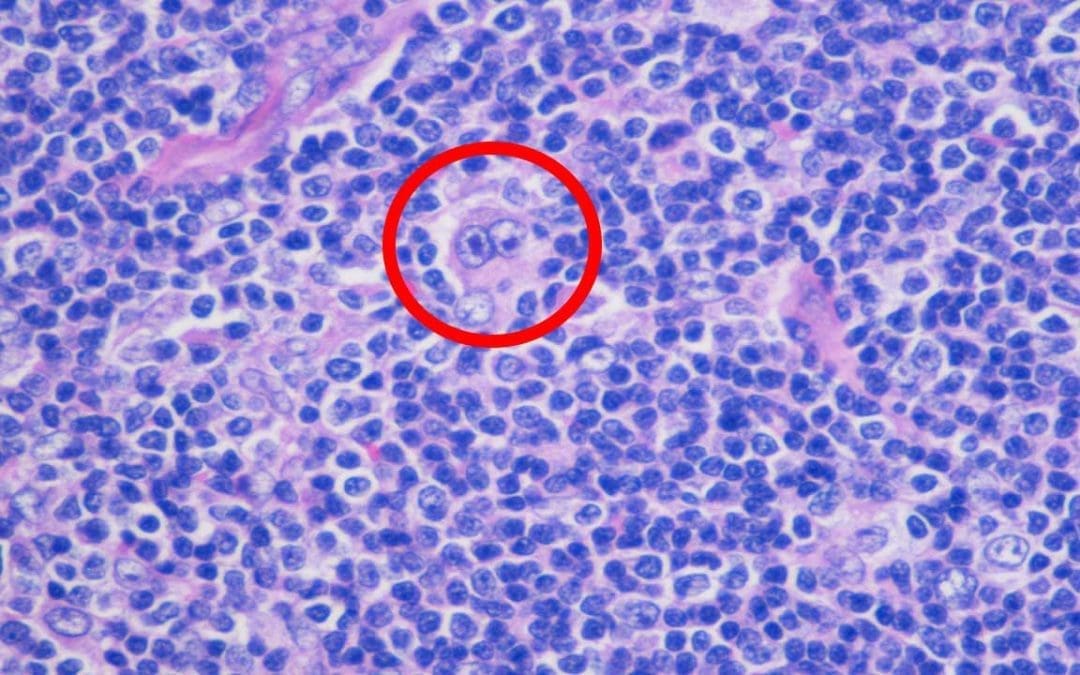
Key Takeaways Hodgkin Lymphoma or formerly known as Hodgkin’s disease is a heterogeneous group of disorders caused by malignant lymphocytes presenting as lymphadenopathy and defined by Reed-Sternberg cells. Hodgkin lymphoma signs and symptoms ▾: The most...
by MyH Editorial Team | Oct 9, 2023 | White Blood Cells
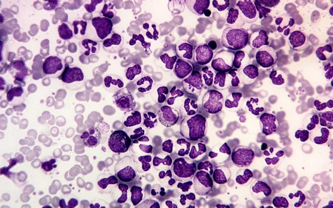
Key Takeaways Chronic myeloid leukemia is a slow-growing blood cancer caused by the BCR-ABL1 fusion gene on the Philadelphia chromosome, formed by a swap between chromosomes 9 and 22. Pathogenesis ▾: Signs and symptoms ▾: Most patients are diagnosed in...
by MyH Editorial Team | Oct 9, 2023 | White Blood Cells
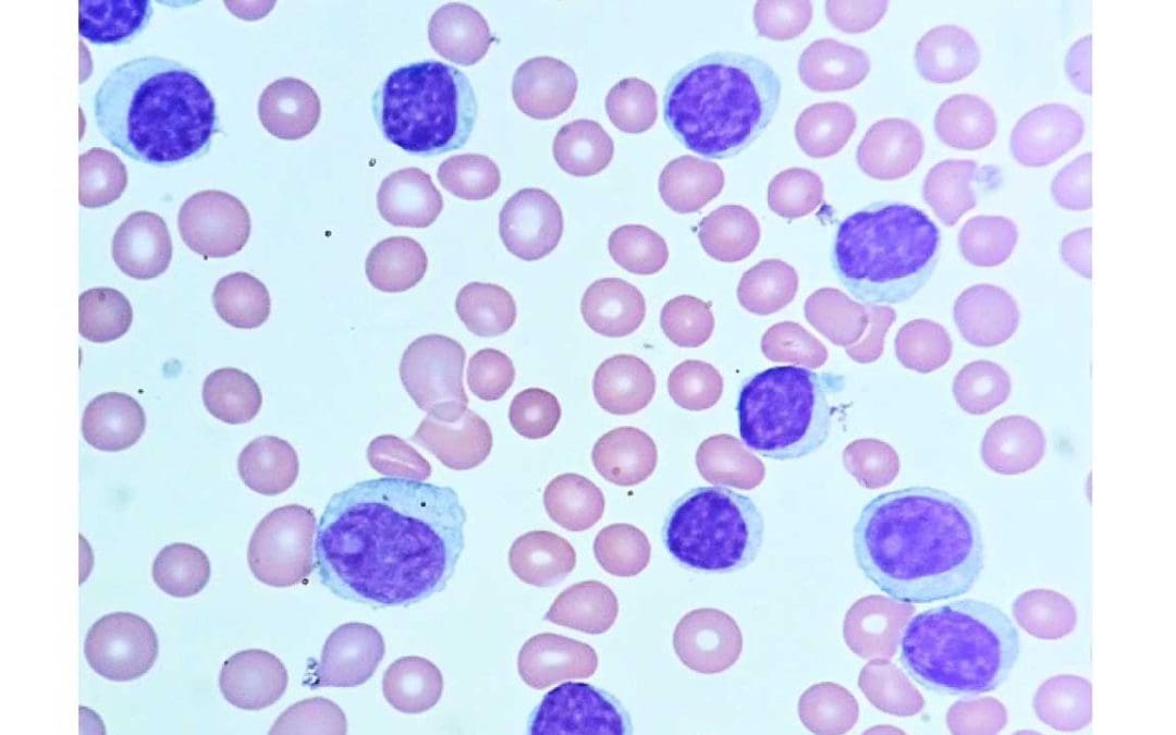
Key Takeaways Chronic lymphocytic leukemia also known as CLL is a slow progressing cancer due to overgrowth and accumulation of small incompetent mature-looking B-lymphocytes in the blood, bone marrow and lymphoid tissues. Small lymphocytic leukemia is a different...
by MyH Editorial Team | Oct 9, 2023 | White Blood Cells
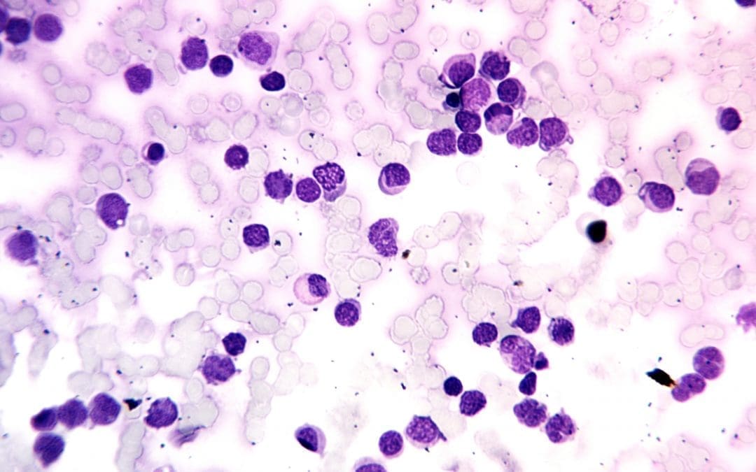
Key Takeaways Acute leukemia is a fast-progressing bone marrow cancer in which immature cells called blasts crowd out normal blood production, causing anemia, infections, and bleeding within days to weeks. AML and ALL differ by cell lineage: AML arises from myeloid...

Recent Comments