Procedure At-A-Glance
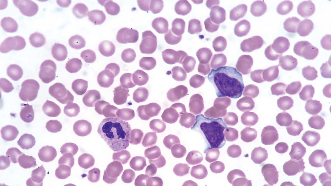
The Wright-Giemsa stain is a combination Romanowsky-type stain used to differentiate blood and bone marrow cell types with more intense nuclear detail than Wright stain alone.
Flood Slide Protocol
- Air-dry a well-made smear. Fix in methanol.
- Flood the slide with undiluted Wright-Giemsa stain. Wait 2–3 minutes.
- Add an equal or greater volume of buffer (pH 6.8). A green metallic sheen confirms correct mixing.
- Stain for 5–15 minutes (bone marrow needs longer than blood films).
- Rinse with buffer or distilled water until edges turn pinkish-red.
- Air-dry the slide.
- Examine under oil immersion.
Dip Slide Protocol with Coplin Jars
- Fix the smear in absolute methanol for 15–30 seconds.
- Dip in Wright-Giemsa stain for 2–3 minutes.
- Transfer to buffer/stain mix (1:5 to 1:10 dilution, pH 6.8) for 5–15 minutes.
- Rinse in distilled water until edges are pinkish-red.
- Air-dry and examine.
Introduction
When a bone marrow biopsy or a peripheral blood smear needs the clearest possible view of nuclear detail, most hematology laboratories reach for the Wright-Giemsa stain. This combination stain merges the speed of Wright stain with the richer basophilic staining of Giemsa, making it the standard choice for both routine blood films and diagnostic bone marrow work. This guide explains what Wright-Giemsa stain is, how it works, how to prepare and apply it step-by-step, and how to interpret what it reveals.
Why Wright-Giemsa Is the Standard Combination Stain
Wright stain alone is fast and reliable for routine differential counts. Giemsa stain alone gives rich nuclear detail but takes longer and is better suited to thick films. Wright-Giemsa stain combines both, giving laboratories a single reagent that performs well on peripheral blood films and produces the intense basophilic and nuclear staining needed to interpret bone marrow aspirates [3].
Wright-Giemsa stain is used across several settings:
- Routine hematology, to evaluate red cells, white cells, and platelets when a blood count is abnormal.
- Bone marrow assessment, where its stronger nuclear contrast helps identify blast cells, abnormal chromatin patterns, and cytoplasmic granularity.
- Parasitology, for rapid detection of blood parasites such as Plasmodium and Trypanosoma species when quick results are needed [1].
Once stained, a smear can reveal immature cells, abnormal nuclear shapes, and cytoplasmic inclusions that point toward conditions such as leukemia, lymphoma, or anemia.
If you are new to blood films, start with our companion article on Preparation of Peripheral Blood Smears. A poorly made smear cannot be rescued by even the best stain.
When Is Wright-Giemsa Stain Indicated?
Wright-Giemsa stain is typically chosen, rather than Wright stain alone, when:
- A bone marrow aspirate or biopsy needs staining, since Wright-Giemsa gives clearer nuclear chromatin patterns for identifying blast cells and abnormal maturation.
- A peripheral smear shows possible blasts or abnormal cells that need closer nuclear evaluation.
- Plasma cell disorders such as multiple myeloma are suspected, since Wright-Giemsa clearly shows the eccentric nucleus, perinuclear halo, and clock-face chromatin characteristic of plasma cells.
- Parasites need rapid identification in blood or bone marrow.
Principle of the Wright-Giemsa Stain
Wright-Giemsa belongs to the Romanowsky family of stains. Its name reflects its origin: the Romanowsky principle traces back to Dmitri Romanowsky's 1891 observation that combining oxidized methylene blue (azure dyes) with eosin produced a distinctive purple color unattainable with either dye alone [1]. Over the following decades, various chemists, including Wright and Giemsa, refined this combination, and Wright-Giemsa stain represents a further blending of their two formulations.
The stain works through the same basic chemistry as its parent stains:
- Azure dyes (oxidized methylene blue), positively charged, bind negatively charged nucleic acids in the nucleus and RNA-rich cytoplasm, producing blue to purple coloration.
- Eosin Y, negatively charged, binds positively charged proteins such as hemoglobin, producing pink to red coloration.
The Giemsa component adds additional azure dyes not present in Wright stain alone, which increases the intensity of nuclear and basophilic cytoplasmic staining. This is the main practical reason laboratories choose Wright-Giemsa over Wright stain when nuclear detail matters most, such as in bone marrow interpretation [1, 3].
As with Wright stain, the methanol solvent fixes cells to the slide, and the buffer, typically pH 6.8, triggers the staining reaction and controls the final color balance. Distilled or deionized water should be used for buffer and rinse steps, since variation in tap water pH can affect results [5].
Method 1: Flood Slide Technique
Materials
- Wright-Giemsa stain solution (commercial combination stain, methanol-based)
- Phosphate buffer, pH 6.8 (Sorensen's or equivalent)
- Staining rack
- Pasteur pipette
- Absolute methanol (fixative)
- Timer
- Distilled or deionized water for rinsing
- Microscope with oil immersion
Protocol
- Prepare a well-made smear (blood or bone marrow) and allow it to air-dry completely.
- Fix in absolute methanol for 15–30 seconds, then allow to air-dry.
- Flood the slide with filtered, undiluted Wright-Giemsa stain. Let it stand for 2–3 minutes.
- Add an equal or greater volume of buffer (pH 6.8). Mix gently by blowing across the surface. A green metallic sheen signals correct mixing.
- Stain for 5–15 minutes. Peripheral blood films need less time; bone marrow smears typically need two to three times longer than blood films [4].
- Rinse with buffer or distilled water until the thin edges of the smear turn pinkish-red.
- Air-dry at room temperature.
- Examine first on low power, then under oil immersion.
Method 2: Dip Slide Technique with Coplin Jars
Materials
- Wright-Giemsa stain solution
- Phosphate buffer, pH 6.8
- Absolute methanol
- 4 Coplin jars
- Forceps, timer
- Distilled or deionized water
Protocol
- Fill the Coplin jars: methanol, undiluted Wright-Giemsa stain, buffer/stain mix, and distilled water, respectively.
- Fix smears in absolute methanol for 15–30 seconds.
- Submerge in undiluted stain for 2–3 minutes.
- Transfer to the buffer/stain mix (commonly diluted 1:5 to 1:10 with buffer) for 5–15 minutes. This is where most of the staining takes place [3].
- Rinse in distilled water until the edges turn pinkish-red.
- Air-dry and examine.
Practical note
Consistency matters more than speed. Small variations in fixation time, buffer pH, or rinse water quality accumulate into visible differences in staining quality, so many labs standardize every step tightly rather than adjusting on the fly [4].
Quality Control and Modern Practice
Routine QC steps for Wright-Giemsa stain include:
- Check buffer pH daily. Distilled or deionized water is preferred over tap water because of pH variability [5].
- Run a normal control smear alongside patient smears, especially bone marrow controls, to confirm consistent staining.
- Filter the stain before use to prevent fine precipitates.
- Many manufacturers recommend discarding working Wright-Giemsa stain after each use rather than reusing it, since dye stability in an open working solution is limited [5].
Modern hematology laboratories increasingly use digital cell-imaging analyzers for peripheral blood, though bone marrow interpretation still relies heavily on manual microscopy of Wright-Giemsa stained smears. Consistent staining remains essential for both. The International Council for Standardization in Haematology (ICSH) publishes standardized morphology nomenclature that applies regardless of which Romanowsky stain variant is used [7].
Interpretation
| Cell Type | Nucleus | Cytoplasm | Granules | Key Features |
|---|---|---|---|---|
| Red blood cells | None | Pink to salmon | None | Biconcave disc with central pallor |
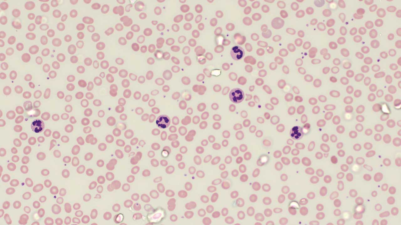
| Neutrophils | Dark purple, 2–5 lobes | Pale pink | Fine lilac | Segmented mature form |
| Eosinophils | Blue, bilobed | Blue | Coarse, bright red-orange | Distinctive large granules |
| Basophils | Dark, often obscured | Pale blue | Coarse, deep purple to black | Granules dominate the cell |
| Monocytes | Blue-violet, kidney or horseshoe shape | Grey-blue, vacuolated | Fine azurophilic | Largest white cell |
| Lymphocytes | Round, dense, dark purple | Sky-blue, scant | Usually none | Small cells, thin cytoplasm rim |
| Plasma cells | Eccentric, clock-face chromatin | Deep basophilic, perinuclear halo | None | Marker of plasma cell disorders such as myeloma [6] |
| Megakaryocytes | Multilobed, large | Abundant, granular | Fine azurophilic | Large marrow cell, platelet precursor |
| Blasts | Fine chromatin, prominent nucleoli | Variable basophilia | Variable; Auer rods in some myeloblasts | Immature cell, high nucleus-to-cytoplasm ratio |
The clock-face chromatin and perinuclear halo of plasma cells are a well-documented example of how Wright-Giemsa's stronger nuclear staining aids diagnosis, as illustrated in bone marrow evaluations of multiple myeloma [6]. Similarly, Auer rods in acute promyelocytic leukemia can be visualized with Wright-Giemsa stain, though comparative research has found their detection rate and clarity can vary between Romanowsky stain variants, an important consideration when a diagnosis hinges on this finding [5].
Troubleshooting
Staining technique
| Problem | Common causes | Solutions |
|---|---|---|
| Too blue (excess basophilia) | Alkaline buffer or rinse water, prolonged staining, thick smear | Adjust buffer pH toward 6.5–6.8, shorten stain time, use thinner smears |
| Too pink (excess eosinophilia) | Acidic buffer, prolonged washing, understaining | Adjust buffer pH, reduce wash time, extend stain time |
| Weak nuclear detail on bone marrow | Insufficient stain time, using Wright stain time on marrow smears | Extend staining to 2–3 times the blood film time [4] |
| Precipitate on slide | Unfiltered stain, tap water used for rinsing | Filter stain before use, switch to distilled or deionized water [5] |
| Poor Auer rod visibility | Stain variant and preparation differences | Consider alternate stains such as Liu's stain if Auer rod detection is diagnostically critical [5] |
| Inconsistent day-to-day results | Variation in fixation time, buffer pH, or technique | Standardize every step; small variations accumulate into visible differences [4] |
Comparison with Other Romanowsky Stains
| Stain | Key features | Advantages | Disadvantages |
|---|---|---|---|
| Wright-Giemsa | Combined Wright and Giemsa azures; 5–15 min | Strong nuclear detail, ideal for bone marrow and peripheral blood, versatile | Slower than Wright alone; requires careful standardization |
| Wright | Methylene blue + eosin alone; 3–5 min | Fast, reliable for routine differential counts | Less nuclear detail than Wright-Giemsa |
| Giemsa | Pure azures + Eosin Y; ~30 min, glycerol-based | Gold standard for thick films and malaria parasite detection | Long preparation and staining time |
| Liu's stain | Rapid modified Romanowsky stain | Superior detection of Auer rods and faggot cells in some studies [5] | Less widely available outside parts of Asia |
| Leishman | Polychromed methylene blue + eosin; 15–20 min | Simple, good host-cell contrast in malaria thin films | Less vivid nuclear detail than Wright-Giemsa |
For routine peripheral blood work, plain Wright stain is often sufficient. When bone marrow interpretation, plasma cell morphology, or the clearest possible nuclear detail is needed, Wright-Giemsa stain is generally the preferred combination [1,3].
Safety and Waste Disposal
Wright-Giemsa stain contains methanol, which is flammable and toxic if inhaled, swallowed, or absorbed through the skin. Practical precautions:
- Wear gloves and eye protection. Work in a ventilated space or fume hood.
- Keep stain bottles away from open flames and sparks.
- Collect used stain and rinses in labeled flammable hazardous-waste containers. Do not pour stain waste down the drain or evaporate it.
- Follow your institution's chemical hygiene plan for disposal documentation.
- Refer to the safety data sheet (SDS) provided by your stain manufacturer for full health and safety information [5].
Frequently Asked Questions (FAQs)
What is Wright-Giemsa stain used for?
Wright-Giemsa stain is a combination Romanowsky-type stain used to color peripheral blood and bone marrow smears for microscopic examination. It is especially valued for bone marrow interpretation because it produces stronger nuclear and basophilic detail than Wright stain alone.
How is Wright-Giemsa stain different from Wright stain?
Wright-Giemsa combines the Wright stain formulation with additional Giemsa azure dyes. This produces more intense nuclear staining, making it better suited to bone marrow aspirates and cases where fine chromatin detail matters, such as identifying blast cells or plasma cells.
Why does bone marrow need longer staining time than peripheral blood?
Bone marrow smears are typically thicker and contain a higher density of nucleated cells than peripheral blood films. Bone marrow smears generally require two to three times the staining time of a peripheral blood film to achieve adequate dye penetration and contrast [4].
What does the green metallic sheen during staining indicate?
It is a visual cue that the stain and buffer have mixed in the correct proportion. If the sheen does not appear, more buffer is usually needed.
Can Wright-Giemsa stain detect malaria parasites?
Yes. Wright-Giemsa stain can be used for rapid detection of blood parasites such as Plasmodium and Trypanosomaspecies when quick results are needed, although pure Giemsa stain remains the gold standard for thick-film malaria diagnosis [1].
Is Wright-Giemsa stain safe to use?
The dye itself is generally safe under normal laboratory conditions, but methanol, its solvent, is flammable and toxic if inhaled, swallowed, or absorbed through skin. Always wear gloves and eye protection, work in a ventilated area, and dispose of waste through your institution's flammable hazardous-waste stream.
Glossary of Related Medical Terms
- Peripheral blood smear (PBS): A thin film of blood spread on a glass slide, stained, and examined under a microscope.
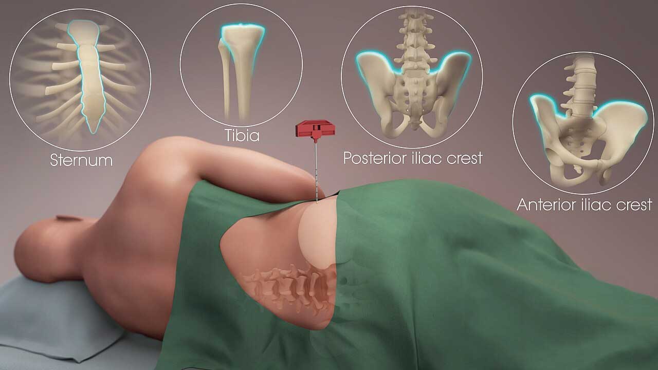
- Bone marrow aspirate: A liquid sample of bone marrow cells drawn by needle, smeared on a slide, and stained to evaluate blood cell production at its source.
- Romanowsky stain: A family of blood stains that combine a basic dye (methylene blue or its azure derivatives) with an acidic dye (eosin) to color cell components in contrasting shades. Wright, Giemsa, Leishman, and Wright-Giemsa stains all belong to this family.
- Azure dyes: Basic, positively charged dyes formed by oxidizing methylene blue. They bind negatively charged nucleic acids, staining nuclei blue to purple.
- Eosin Y: An acidic, negatively charged dye that binds positively charged proteins such as hemoglobin, giving red blood cells their pink to salmon color.
- Polychromatic stain: A stain containing more than one dye component, producing a range of colors rather than a single shade.
- Metallic sheen: A green, film-like reflection on the slide surface that signals stain and buffer have mixed in the correct proportion.
- Fixation: A step that preserves cell structure on the slide, usually with methanol, before staining begins.
- Basophilia (staining context): Increased blue-purple coloration of a cell or smear, caused by stronger binding of the basic azure dyes.
- Eosinophilia (staining context): Increased pink-red coloration of a cell or smear, caused by stronger binding of eosin. (Not to be confused with an elevated eosinophil count in a blood test.)
- Nuclear chromatin: The DNA and protein material inside a cell's nucleus. Its pattern, coarse, fine, or clumped, helps identify cell maturity and type.
- Clock-face chromatin: A chromatin pattern in plasma cells where the nuclear material is arranged in a radial, wheel-like pattern, a classic feature seen with Wright-Giemsa stain.
- Perinuclear halo: A pale, clear zone surrounding the nucleus of a plasma cell, caused by a well-developed Golgi apparatus.
- Plasma cell: A mature white blood cell that produces antibodies. Abnormal proliferation of plasma cells is seen in multiple myeloma.
- Megakaryocyte: A large bone marrow cell responsible for producing platelets.
- Blast cell: An immature precursor cell, normally confined to the bone marrow. Its presence in a peripheral smear or in excess in the marrow can indicate leukemia.
- Auer rod: A needle-shaped, pink to red staining structure in the cytoplasm of some myeloblasts, a key diagnostic clue for acute myeloid leukemia, including acute promyelocytic leukemia.
- Faggot cell: A leukemic promyelocyte containing bundles of multiple Auer rods, a distinctive finding in acute promyelocytic leukemia.
- Nucleus-to-cytoplasm (N:C) ratio: The proportion of a cell's nucleus size to its cytoplasm size. Immature cells like blasts typically have a high N:C ratio.
- Granulocyte: A white blood cell containing visible cytoplasmic granules, including neutrophils, eosinophils, and basophils.
- Thrombocytopenia: A low platelet count.
- Erythropoiesis: The process of red blood cell production in the bone marrow.
Disclaimer: This protocol is for educational purposes only. Local laboratory standard operating procedures take precedence. It is not intended to be a substitute for informed professional medical advice, diagnosis, or treatment. Always consult a qualified healthcare professional for clinical decision-making. While the information presented here is derived from credible medical sources and is believed to be accurate and up-to-date, it is not guaranteed to be complete or error-free. See additional information.
References
- Hardy Diagnostics. (2025). How to do a Wright-Giemsa stain [Technical bulletin]. Retrieved from manufacturer documentation.
- Ward, P., Glassy, E., Kroft, S., & Krafts, K. (2011). A Mysterious Malady: the Malachowski-Wright-Giemsa Stain to the Rescue. Biotechnic & Histochemistry, 86(2), 76–81. https://doi.org/10.3109/10520295.2010.515495
- Dunning, K., & Safo, A. O. (2011). The ultimate Wright-Giemsa stain: 60 years in the making. Biotechnic & histochemistry : official publication of the Biological Stain Commission, 86(2), 69–75. https://doi.org/10.3109/10520295.2010.515496
- Thermo Fisher Scientific. (n.d.). Richard-Allan Scientific Wright-Giemsa Stain Solution [Instructions for use, IS89013]. Thermo Fisher Scientific.
- Yue, Q. F., Xiong, B., Chen, W. X., & Liu, X. Y. (2014). Comparative study of the efficacy of Wright-Giemsa stain and Liu's stain in the detection of Auer rods in acute promyelocytic leukemia. Acta histochemica, 116(6), 1113–1116. https://doi.org/10.1016/j.acthis.2014.05.005
- Bain, B. J. & Leach, M. (2025). Blood Cells: A Practical Guide (7th ed.). Wiley.
- ICSH reference method for staining of blood and bone marrow films by azure B and eosin Y (Romanowsky stain). International Committee for Standardization in Haematology. (1984). British journal of haematology, 57(4), 707–710. https://doi.org/10.1111/j.1365-2141.1984.tb02949.x