
by MH Team | Oct 18, 2023 | Transfusion Medicine
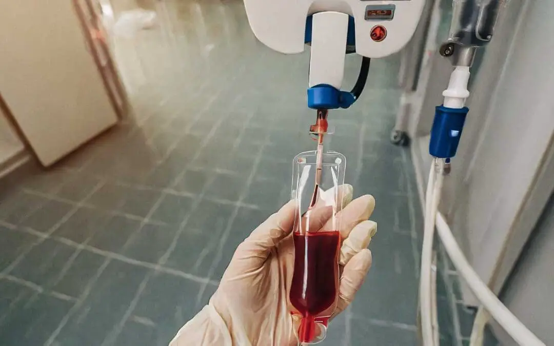
TL;DR Acute Hemolytic Transfusion Reaction (AHTR) is an acute hemolysis due to the administration of antigenically incompatible red cells. Timing ▾: During transfusion or within 24 hours. Signs and Symptoms ▾: Fever and chills Back or flank pain...
by MH Team | Oct 17, 2023 | White Blood Cells
TL;DR Multiple myeloma is a cancer of plasma cells, a type of white blood cell that produces antibodies. In this condition, abnormal plasma cells multiply uncontrollably and accumulate in the bone marrow. It is slightly more common in males than females and most...
by MH Team | Oct 17, 2023 | Platelet Disorders
TL;DR Hemophilia, the most common severe, inherited bleeding disorder, represents a failure in the secondary hemostatic mechanism. It is an X-linked recessive disorder characterized by a deficiency or dysfunction of a specific plasma coagulation factor, leading to...
by MH Team | Oct 16, 2023 | Platelet Disorders
TL;DR Vitamin K Deficiency Bleeding (VKDB) is a hemorrhagic disorder in newborns and infants (typically 0-6 months) caused by insufficient levels of vitamin K-dependent clotting factors, leading to spontaneous or traumatic bleeding. Causes: All newborns are...
by MH Team | Oct 16, 2023 | Platelet Disorders
TL;DR Von Willebrand Disorder also known as Von Willebrand Disease or VWD is an inherited coagulation disorder caused by mutations in the Von Willebrand Factor (VWF). VWF is a factor VIII carrier protein and mediates platelet adhesion to the endothelium. Pathogenesis...


Recent Comments