
by MH Team | Nov 14, 2023 | Red Blood Cells
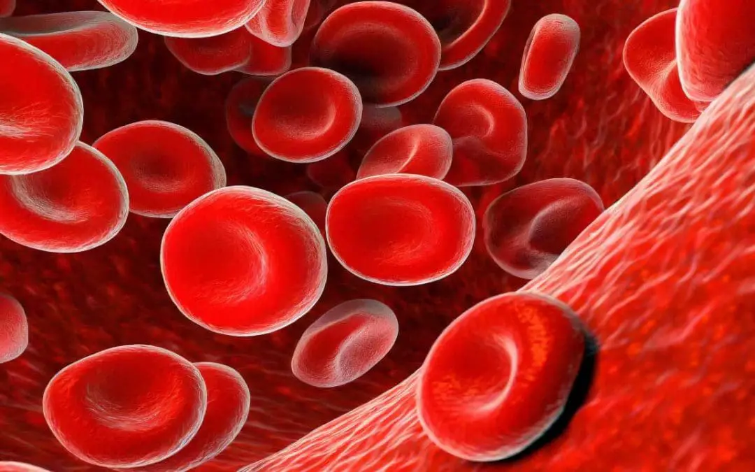
TL;DR Red blood cells (RBCs), or erythrocytes, are the most common blood cells, small biconcave disks lacking a nucleus and primarily composed of hemoglobin. Function ▾: Their main function is to transport oxygen from the lungs to tissues and carbon dioxide...
by MH Team | Nov 14, 2023 | Lab Protocols, Red Blood Cells
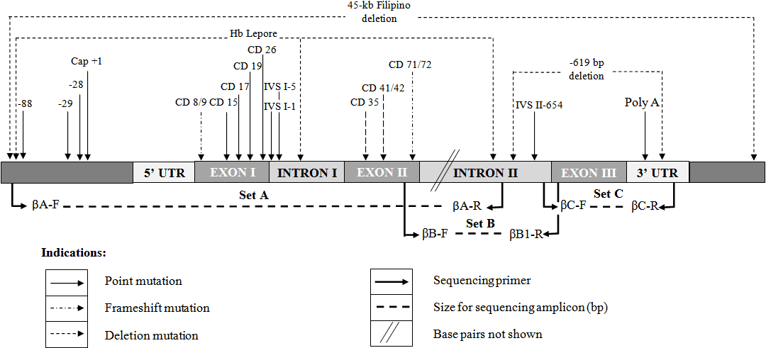
Procedure-at-a-Glance StageActivity1. PreparationReagent thawing & Workstation UV sterilization2. Master MixCombining Polymerase, dNTPs, Buffers, and Primers3. Amplification30 cycles of Denaturation, Annealing, and Extension4. VerificationGel Electrophoresis (1%...
by MH Team | Nov 13, 2023 | Transfusion Medicine
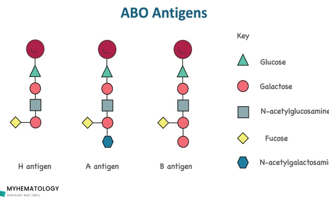
TL;DR ABO blood group is the most important blood group system in transfusion and organ transplantation medicine. ABO blood group antigens can be found on red cells, white cells, platelets and many circulating proteins. Transfusion incompatibility can cause...
by MH Team | Nov 9, 2023 | Lab Protocols, White Blood Cells
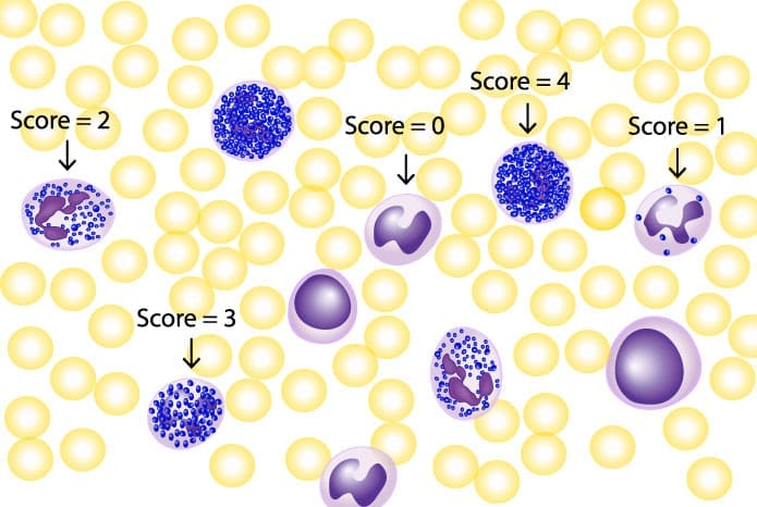
Procedure-at-a-Glance StepActionDuration1. FixationFix fresh smears in Cold Formalin-Methanol (0-10°C).30 Seconds2. WashingGently rinse with running tap water and air dry.—3. IncubationImmerse in freshly prepared buffered substrate.15–30 Minutes4....
by MH Team | Nov 9, 2023 | Red Blood Cells
TL;DR Aplastic anemia (AA) causes pancytopenia (reduction of all blood cell types: RBCs, WBCs and platelets) due to aplasia (failure) of the bone marrow to function (to produce blood cells). Causes▾: Congenital e.g. Fanconi anemia, dyskeratosis congenita...

Recent Comments