Key Takeaways
WBCs, also called leukocytes, are the body's defense cells. They protect against bacteria, viruses, fungi, parasites, and abnormal cells like cancer cells.
- Types of White Blood Cells ▾: There are five main types of WBCs in circulation: neutrophils, lymphocytes, monocytes, eosinophils, and basophils. Each has a distinct role
- White Blood Cells (WBCs) Disorders ▾: Disorders of WBCs range from benign (reactive leukocytosis, neutropenia) to malignant (acute and chronic leukemias). Modern leukemia treatment is increasingly targeted, guided by genetic testing of the leukemic cells.
*Click ▾ for more information
What are white blood cells?
White blood cells, or WBCs, are the body's frontline defense system. They patrol the blood and lymphatic system looking for anything that does not belong: bacteria, viruses, fungi, parasites, and even cells that have turned cancerous. The name leukocyte comes from the Greek leukos (white) and cyte (cell). [1]
Although they share the same job description, the WBCs are actually a small team with very different specialties. Some are first responders that engulf microbes within minutes. Others remember past infections for decades. Knowing who does what is the key to understanding nearly every blood disorder a student will meet on the wards.
This article walks through the five main WBCs, how they are produced, what their normal values look like, and what happens when they go wrong.
What are the types of white blood cells (WBCs)?

WBCs are split into two groups based on whether their cytoplasm contains visible granules. [1]
Granulocytes — neutrophils, eosinophils, basophils — have prominent granules packed with enzymes and chemical mediators.
Agranulocytes — lymphocytes and monocytes — have either no granules or only fine ones. They tend to be more "thinkers" than "fighters."
Neutrophils
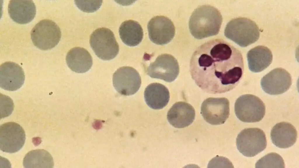
Neutrophils are the most abundant WBCs, making up 40–70% of the total count. They are the first responders to a bacterial or fungal infection, often arriving at the site within hours. [2]
A neutrophil works by chemotaxis (following chemical signals to the infection), engulfing the microbe (phagocytosis), and then releasing a cocktail of enzymes (myeloperoxidase, lysozyme, defensins, lactoferrin) from its granules to kill it. They can also expel webs of DNA studded with antimicrobial proteins called neutrophil extracellular traps (NETs), which trap bacteria. NETs are protective in moderation, but excess NET formation is now linked to thrombosis in sepsis and severe COVID-19. [11]
Under the microscope, a neutrophil has a distinctive 2–5 lobed nucleus and pale pink cytoplasm with very fine granules.
Eosinophils

Eosinophils make up about 1–4% of WBCs. Under the microscope they have a striking bilobed nucleus and bright orange-red granules. Their main jobs are parasite defense and allergic responses. Their granules contain major basic protein, eosinophil cationic protein, and ribonucleases which are substances toxic to parasites.
A persistently raised eosinophil count (eosinophilia) points to allergic disease, parasitic infection, certain drug reactions, or, less commonly, a hematologic disorder.
Basophils
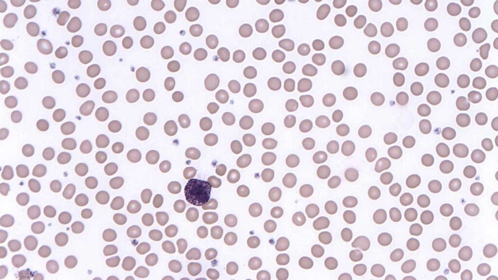
Basophils are the rarest granulocyte at less than 1% of WBCs. Their dark blue-purple granules are so dense they often hide the nucleus. They release histamine, heparin, and IL-4 during allergic reactions. Among circulating leukocytes they are the main histamine carriers, though tissue mast cells (a related but separate cell type) carry far more.
Lymphocytes
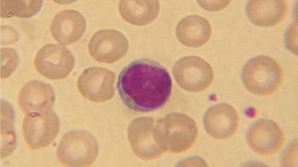
Lymphocytes are the second most abundant WBC and run the adaptive immune response — the slower, specific defense that produces immunological memory [3].
- B cells make antibodies. Once activated by an antigen, they mature into plasma cells that pump out specific antibodies, neutralizing pathogens or marking them for other immune cells to destroy. A subset becomes long-lived memory B cells.
- T cells mature in the thymus and split into helper T cells (which orchestrate the response by activating other immune cells), cytotoxic T cells (which directly kill infected or cancerous cells), and regulatory T cells (which dampen the response so it does not attack the body itself).
- Natural killer (NK) cells sit at the border of innate and adaptive immunity. They kill virus-infected and tumor cells without needing prior exposure, by reading the balance of activating and inhibitory signals on the target cell.
Monocytes
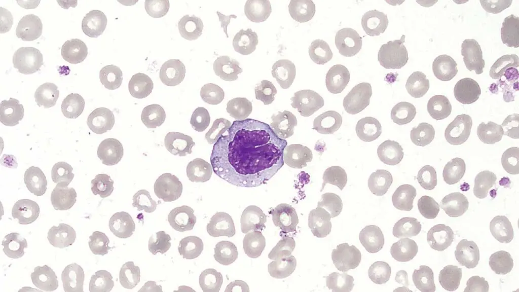
Monocytes are the largest WBCs and have a kidney-shaped nucleus. They circulate for only a few days before migrating into tissues, where they mature into macrophages or dendritic cells (DCs).
Macrophages engulf debris, dead cells, and microbes. They are also the main antigen-presenting cells (APCs) in many tissues, displaying fragments of what they have eaten on their surface to T cells. Tissue-resident macrophages have specific names depending on where they live: alveolar macrophages in the lungs, Kupffer cells in the liver, microglia in the brain, osteoclasts in bone.
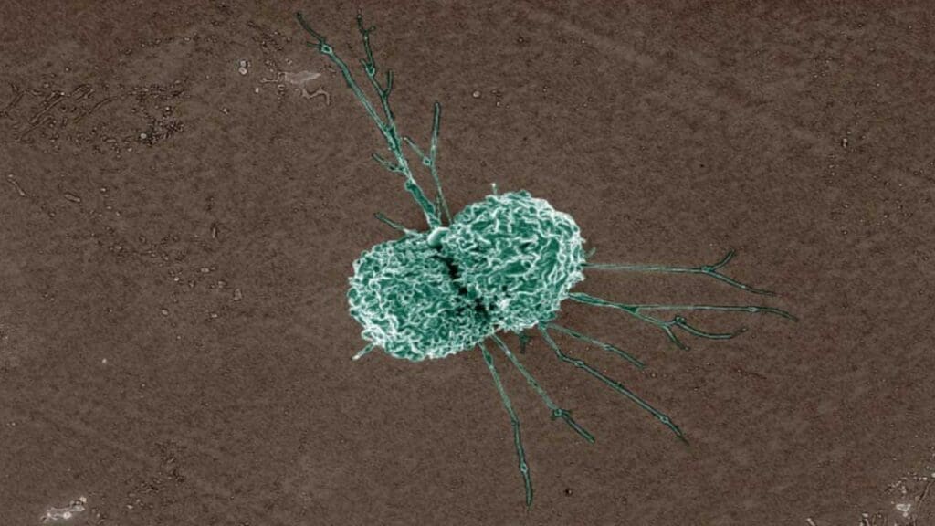
Dendritic cells are the most powerful APCs in the body. They capture antigens in tissues like the skin and gut, then migrate to lymph nodes to "introduce" the antigen to T cells, kicking off the adaptive response.
How WBCs are made: leukopoiesis
All blood cells, including the WBCs, come from a small population of hematopoietic stem cells (HSCs) that live mainly in the bone marrow. The whole process of making blood cells is called hematopoiesis, and the part that makes WBCs is leukopoiesis. [1,13]
A simplified family tree looks like this:
- HSC → can self-renew or commit to a lineage.
- Common myeloid progenitor (CMP) → gives rise to neutrophils, eosinophils, basophils, monocytes, dendritic cells, red cells, and platelets.
- Common lymphoid progenitor (CLP) → gives rise to B cells, T cells, and NK cells.
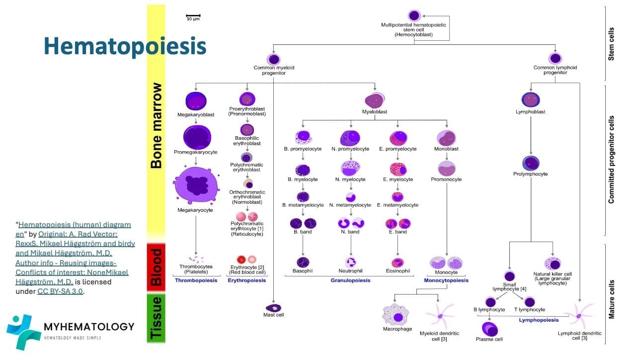
This commitment is steered by cytokines and growth factors. The most important for the WBC lineages are:
- G-CSF — drives neutrophil production. Used clinically as filgrastim or pegfilgrastim to boost counts after chemotherapy.
- GM-CSF — supports granulocyte and monocyte development.
- IL-3 — supports many myeloid lineages.
- IL-5 — drives eosinophil production.
- IL-7 — essential for B and T cell development.
- SCF (stem cell factor) and Flt3-ligand — keep stem cells alive and dividing.
Granulopoiesis
Neutrophils, eosinophils, and basophils all develop through the same series of stages, taking roughly 7–14 days end to end:
Myeloblast → promyelocyte → myelocyte → metamyelocyte → band → segmented mature granulocyte.
The earliest stages are driven by SCF, IL-3, and GM-CSF. From the myelocyte stage onward, lineage-specific cytokines take over: G-CSF for neutrophils, IL-5 for eosinophils, IL-3 plus SCF for basophils.
Lymphopoiesis
B cells start and finish in the bone marrow. They progress through pro-B → pre-B → immature B stages, learning along the way to recognize foreign antigens but not the body's own tissues as a quality control step called self-tolerance.
T cells start in the bone marrow but finish in the thymus, where they undergo a rigorous two-step quality check: positive selection (must be able to recognize self-MHC) and negative selection (must not bind too strongly to self-antigens). Most thymocytes fail this gauntlet and die. The survivors emerge as either CD4+ helper T cells or CD8+ cytotoxic T cells.
How WBC numbers are kept in check
When an infection takes hold, cytokines like IL-1, IL-6, and TNF-α push the bone marrow to make more WBCs, especially neutrophils. As the infection clears, mature neutrophils that are no longer needed are cleared by tissue macrophages, and that clearance reduces G-CSF production thereby turning down WBC output. The spleen also helps, storing mature lymphocytes and releasing or reabsorbing them as needed.
Hormones add another layer. Glucocorticoids (cortisol) raise neutrophil counts by releasing them from the marginated pool but lower lymphocyte counts. This is why a stressed or steroid-treated patient can show a striking neutrophilia on the next CBC.
Normal ranges and the WBC differential
A complete blood count (CBC) with differential is one of the most ordered tests in medicine.
- Total WBC count: 4,000–11,000 cells/µL (4–11 × 10⁹/L).
- Neutrophils: 40–70%
- Lymphocytes: 20–40%
- Monocytes: 2–10%
- Eosinophils: 1–4%
- Basophils: 0.5–1%
Several clinical concepts hang off this differential:
Absolute neutrophil count (ANC) is the actual neutrophil number per µL, calculated from the total WBC and the neutrophil percentage. It is the single most important number for assessing infection risk in chemotherapy patients. An ANC below 0.5 × 10⁹/L is severe neutropenia.
Left shift means the marrow is rushing immature neutrophils (band cells) into the blood, usually because of severe bacterial infection or systemic inflammation. It is a clue, not a diagnosis.
Febrile neutropenia is a medical emergency: fever (≥38.3 °C once, or ≥38.0 °C sustained) with an ANC under 0.5 × 10⁹/L. Patients are admitted and started on broad-spectrum intravenous antibiotics within an hour, before the infecting organism is identified.
Variations that are not disease
Normal does not mean identical. Newborns and young children run higher counts than adults. Pregnancy, smoking, exercise, and even time of day shift the numbers.
One important variation deserves its own paragraph: Duffy-null associated neutrophil count (DANC). Many people of African or Middle Eastern ancestry have lower baseline neutrophil counts than the European-derived reference range suggests. This is caused by a common variant in the ACKR1 gene (the Duffy antigen receptor) that shifts neutrophils into the tissues rather than the blood. Their neutrophils still work normally and they are not at increased risk of infection [4,5]. DANC has historically been called "benign ethnic neutropenia," but that term has been retired. Recent work has proposed a Duffy-null–specific lower limit for the ANC of around 1.21 × 10⁹/L, and major bodies such as the American Society of Hematology now recommend using DANC-aware thresholds in clinical trial eligibility, chemotherapy dosing, and clozapine monitoring. This matters because misclassifying DANC as neutropenia has historically led to unnecessary tests and even withholding of chemotherapy.
WBCs in Inflammation
Inflammation is the body's response to injury, infection, or irritation. Each WBC type plays a different part:
- Neutrophils arrive first, kill microbes by phagocytosis, and release reactive oxygen species and NETs.
- Monocytes follow, mature into macrophages, clean up debris, and start presenting antigens to T cells.
- Lymphocytes mount the targeted, specific response with antibodies from B cells and direct killing or cytokine release from T cells.
- Eosinophils and basophils dominate in allergic and anti-parasitic responses, releasing histamine and other mediators that cause the swelling, itch, and bronchoconstriction of an allergic reaction.
The whole process is coordinated by chemical messengers: cytokines (IL-1, IL-6, TNF-α), chemokines that attract specific cell types, and histamine released by basophils and mast cells.
Acute, short-lived inflammation is healing. Chronic inflammation is harmful as it drives diseases like rheumatoid arthritis, asthma, atherosclerosis, and even some cancers. This is why anti-inflammatory medications, from NSAIDs to biologics targeting specific cytokines, are such a large part of modern therapy.
Benign WBC Disorders
These are non-cancerous conditions where WBC numbers or function are abnormal.
Leukocytosis (WBC > 11,000/µL)
Most often caused by infection, inflammation, stress, pregnancy, or steroid use. Treatment is directed at the cause; the count itself usually does not need correcting. A leukocytosis above 50,000/µL with a left shift, but no underlying leukemia, is called a leukemoid reaction — a dramatic but benign response to a strong stimulus like severe infection or hemorrhage.
Leukopenia (WBC < 4,000/µL)
Causes include:
- Bone marrow problems: aplastic anemia, infiltration by tumor, B12/folate deficiency.
- Increased destruction: autoimmune disease, hypersplenism.
- Drugs: chemotherapy, some antibiotics, antiepileptics, antithyroid drugs.
The clinical worry is infection risk, especially when the ANC drops below 0.5 × 10⁹/L. Patients are taught neutropenic precautions (avoiding crowds, raw foods, gardening), and any fever is treated as an emergency.
Lineage-specific disorders
| Pattern | Common Causes |
|---|---|
| Neutrophilia | Bacterial infection, inflammation, stress, burns, surgery, steroids |
| Neutropenia | Chemotherapy, viral infection, autoimmune (Felty syndrome, SLE), B12/folate deficiency, congenital (cyclic neutropenia, severe congenital neutropenia from ELANE mutations) |
| Eosinophilia | Allergy, parasites, drug reactions, hypereosinophilic syndrome |
| Basophilia | Allergic disorders, chronic myeloid leukemia, polycythemia vera |
| Monocytosis | TB, endocarditis, chronic infections, autoimmune disease, monocytic leukemias |
| Lymphocytosis | Viral infections (especially EBV, CMV), pertussis, CLL |
| Lymphopenia | HIV, steroids, chemotherapy, severe acute illness, congenital immunodeficiency |
Inherited WBC anomalies
A few conditions show up on blood films and exam questions:
- May-Hegglin anomaly — MYH9 mutation, giant platelets, Döhle-like inclusions in neutrophils, mild thrombocytopenia.
- Pelger-Huët anomaly — LBR (lamin B receptor) mutation [12, 13]. Neutrophils have hyposegmented, dumbbell-shaped nuclei but function normally. This is sometimes confused with ELANE-related neutropenia, which is a separate condition.
- Chediak-Higashi syndrome — CHS1/LYST mutation, giant lysosomes, partial albinism, and recurrent infections because neutrophils cannot deliver their granules properly.
Severe burns: a clinical example
Burns trigger a dramatic and biphasic WBC response. In the first hours there is a stress-driven neutrophilia. Over the following days, the marrow's reserves run down and neutrophils are sequestered in damaged tissue, often leaving the patient leukopenic and at high risk of infection. This is a major reason burn patients are nursed in protective isolation.
Malignant WBC Disorders
Leukemias are cancers in which a single abnormal WBC clone takes over the bone marrow, crowding out healthy red cells, platelets, and normal WBCs. They are split by speed (acute versus chronic) and by lineage (myeloid versus lymphoid), giving four main types [10].
Acute Myeloid Leukemia (AML)
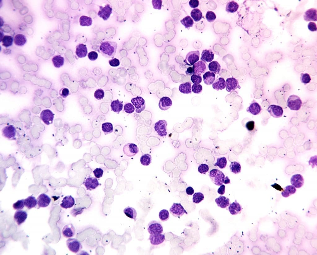
A rapidly progressive cancer of immature myeloid cells (myeloblasts) that flood the marrow and blood.
- Risk factors: prior chemotherapy or radiation, benzene exposure, certain genetic syndromes (Down syndrome, Fanconi anemia).
- Symptoms: fatigue, fever, easy bruising, frequent infections, weight loss.
- Diagnosis: CBC, peripheral smear (often showing blasts and Auer rods), bone marrow biopsy, flow cytometry, cytogenetics, and molecular testing for FLT3, NPM1, IDH1/2, TP53, and other mutations.
- Treatment: for fit patients, intensive "7+3" chemotherapy (cytarabine plus an anthracycline). Treatment is now mutation-driven: midostaurin or quizartinib is added for FLT3-mutated AML [7], gemtuzumab ozogamicin for CD33-positive core-binding-factor AML, and CPX-351 for therapy-related or MDS-related AML. For older or unfit patients, venetoclax + azacitidine has transformed outcomes, with response rates around 65% in patients who could not previously be treated [6]. Ivosidenib (IDH1) and enasidenib (IDH2) are used for IDH-mutated disease. Allogeneic stem cell transplant remains the main curative option for high-risk patients in remission. A major recent breakthrough is the introduction of targeted menin inhibitors for patients with NPM1 mutations or KMT2A rearrangements. Drugs such as revumenib and the once-daily ziftomenib block the menin protein, forcing leukemia cells to differentiate and mature rather than multiply, which has transformed the prognosis for this large subgroup of AML patients [14,15].
Acute Lymphoblastic Leukemia (ALL)
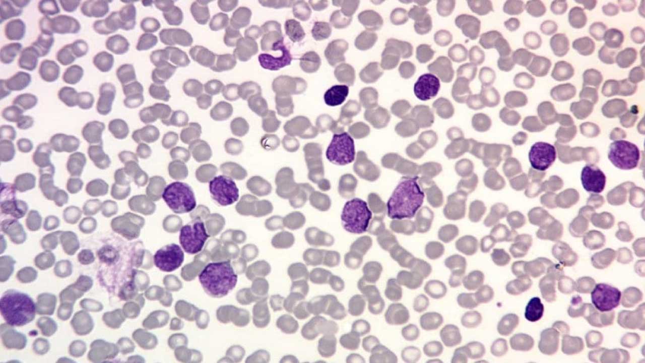
A cancer of immature lymphocytes (lymphoblasts), most commonly B-cell ALL. It is the most common cancer of childhood but also occurs in adults.
- Symptoms: similar to AML, plus bone pain, lymphadenopathy, and CNS involvement (headaches, cranial nerve palsies).
- Diagnosis: as for AML, with subtyping into B-ALL or T-ALL, and testing for the Philadelphia chromosome (BCR-ABL1) and other genetic features that change treatment.
- Treatment: multi-phase chemotherapy with CNS prophylaxis. Major recent additions include blinatumomab (a bispecific T-cell engager that links T cells to CD19 on B-ALL cells), inotuzumab ozogamicin (an anti-CD22 antibody-drug conjugate), and CD19 CAR T-cell therapy (tisagenlecleucel) for relapsed or refractory disease in children and young adults [10]. The CAR T-cell landscape expanded significantly with the late 2024 approval of obecabtagene autoleucel (Aucatzyl) for adults with relapsed or refractory B-ALL. This therapy is uniquely designed to mimic physiological T-cell receptor interactions, resulting in remarkably lower rates of severe cytokine release syndrome (CRS) compared to earlier generations [16]. Tyrosine kinase inhibitors are added for Philadelphia-positive ALL.
Chronic Myeloid Leukemia (CML)
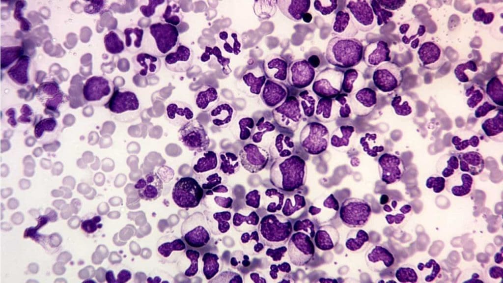
A slow-growing leukemia caused by the Philadelphia chromosome, which is a translocation between chromosomes 9 and 22 that creates the BCR-ABL1 fusion gene. The BCR-ABL1 protein is a constantly-on tyrosine kinase that drives uncontrolled granulocyte proliferation [9].
- Symptoms: often none at diagnosis; sometimes fatigue, weight loss, or abdominal fullness from splenomegaly. The CBC shows a striking leukocytosis with the full granulocyte spectrum, basophilia, and eosinophilia.
- Treatment: the tyrosine kinase inhibitors (TKIs) were the breakthrough. Imatinib was the first; second-generation TKIs include dasatinib, nilotinib, and bosutinib. Ponatinib is used for T315I-mutated disease, and asciminib is a newer agent that targets BCR-ABL1 at a different site. With current TKIs, life expectancy for most patients is now close to that of the general population [9].
Chronic Lymphocytic Leukemia (CLL)
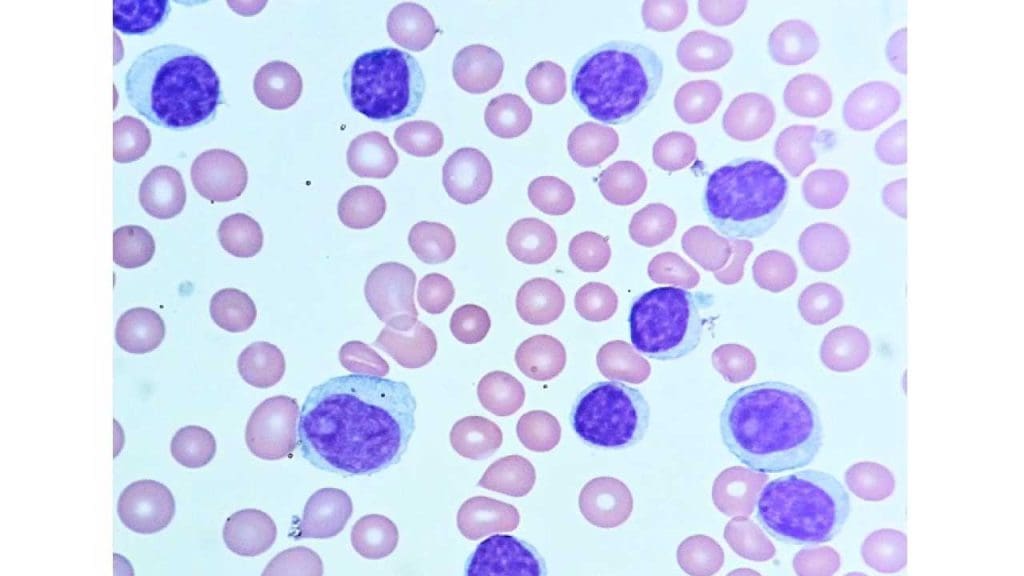
A slow-growing cancer of mature B lymphocytes. Often picked up incidentally on a CBC in older adults.
- Symptoms: often none. As disease progresses: fatigue, lymph node enlargement, recurrent infections, and weight loss.
- Diagnosis: CBC showing lymphocytosis with characteristic small mature lymphocytes (and "smudge cells" on the smear), plus flow cytometry showing the characteristic CD5+/CD19+/CD23+ B-cell phenotype.
- Treatment: chemoimmunotherapy has been completely superseded by targeted agents in current guidelines and is no longer recommended [17]. First-line treatment is targeted: a BTK inhibitor (ibrutinib, acalabrutinib, or zanubrutinib) given continuously, or fixed-duration venetoclax + obinutuzumab [8]. For patients whose disease develops resistance to these older, covalent BTK inhibitors, the non-covalent BTK inhibitor pirtobrutinib is now a licensed and highly effective standard of care [18]. Newer combinations adding BTK inhibitors to venetoclax-based regimens are under active study. Many patients with early-stage disease and no symptoms simply receive "watch and wait". Treatment does not improve outcomes in that group.
How are leukemias diagnosed in practice?
A typical workup combines:
- CBC and peripheral smear — the first hint.
- Bone marrow aspiration and biopsy — confirms diagnosis and gives material for further testing.
- Flow cytometry (immunophenotyping) — uses fluorescent antibodies against CD markers to pin down the lineage and maturation stage of the leukemic cell. Essential for diagnosis and subtyping.
- Cytogenetics and FISH — looks for whole-chromosome changes like the Philadelphia chromosome.
- Molecular testing (NGS) — looks for mutations that determine prognosis and guide targeted therapy.
Frequently Asked Questions (FAQs)
What is a normal WBC count, and what makes it go up or down?
A normal total WBC count in adults is roughly 4,000 to 11,000 cells per microliter. Counts rise (leukocytosis) with infection, inflammation, stress, pregnancy, smoking, exercise, and certain leukemias. Counts fall (leukopenia) with bone marrow problems, chemotherapy, autoimmune destruction, severe infections, or some inherited conditions. A single abnormal value is rarely diagnostic on its own; doctors look at the full differential and the clinical picture.
What is the difference between leukemia and lymphoma?
Leukemia starts in the bone marrow and pours abnormal WBCs into the bloodstream. Lymphoma starts in lymph nodes, the spleen, or other lymphoid tissue and tends to form solid tumors there. Leukemias are typically diagnosed by blood tests and bone marrow biopsy; lymphomas are usually diagnosed by lymph node biopsy. Both are cancers of WBCs but they behave differently and are treated differently.
Why do some healthy people have a low neutrophil count?
Some people, especially those of African or Middle Eastern ancestry, have a genetic variant called the Duffy-null genotype (in the ACKR1 gene) that produces a naturally lower circulating neutrophil count. This is now called Duffy-null associated neutrophil count, or DANC. People with DANC are not at increased risk of infection as their neutrophils still work normally; they just spend more time in the tissues than in the blood. [4,5]
What is febrile neutropenia and why is it an emergency?
Febrile neutropenia is fever in a patient whose neutrophil count has dropped, usually below 0.5 × 10⁹/L, often after chemotherapy. It is an emergency because the patient cannot mount a normal inflammatory response, and a small infection can become life-threatening within hours. Treatment is immediate broad-spectrum intravenous antibiotics, even before the source is identified. Chemotherapy patients are usually told to attend hospital for any temperature above 38 °C.
What does a "left shift" mean on a blood report?
A left shift means the bone marrow is releasing immature neutrophils (band cells, and occasionally metamyelocytes) into the bloodstream. This usually points to a serious bacterial infection, severe inflammation, or major physiological stress like burns or hemorrhage. It can also be seen in chronic myeloid leukemia, where the immature cell mix is more extreme and persistent.
How are leukemias diagnosed today?
Diagnosis usually starts with a complete blood count and a peripheral blood smear. If leukemia is suspected, the next steps are bone marrow aspiration and biopsy, flow cytometry to identify the lineage and maturation stage of the abnormal cells, cytogenetics to look for chromosomal changes like the Philadelphia chromosome, and molecular testing for mutations such as FLT3, IDH1/2, NPM1, and BCR–ABL1. These results guide the choice of targeted therapy.
Glossary of Related Medical Terms
- Leukocyte: Another word for white blood cell.
- Hematopoiesis: The continuous production of blood cells in the bone marrow.
- Leukopoiesis: The part of hematopoiesis that makes WBCs.
- Phagocytosis: A cell engulfing and digesting microbes or debris.
- Cytokine: A small signaling protein immune cells use to communicate.
- Antigen: A molecule (usually on a microbe) the immune system recognizes as foreign.
- Antibody: A Y-shaped protein made by B cells that binds a specific antigen.
- Innate immunity: The fast, non-specific arm of the immune system.
- Adaptive immunity: The slower, specific arm that produces immunological memory.
- Absolute neutrophil count (ANC): Neutrophils per microliter of blood; the key infection-risk number.
- Left shift: Increase in immature neutrophils (band cells) on a blood film.
- Febrile neutropenia: Fever plus an ANC < 0.5 × 10⁹/L; a medical emergency.
- Leukocytosis / Leukopenia: High or low total WBC count.
- Leukemia: Cancer arising in bone marrow that produces abnormal WBCs.
- Lymphoma: Cancer of mature lymphocytes arising in lymph nodes or other lymphoid tissue.
- DANC: Duffy-null associated neutrophil count — a normal, inherited lower neutrophil count.
- CBC with differential: The blood test that measures total WBCs and the percentage of each type.
Disclaimer: This article is intended for educational and informational purposes only. It is not intended to be a substitute for informed professional medical advice, diagnosis, or treatment. While the information presented here is derived from credible medical sources and is believed to be accurate and up-to-date, it is not guaranteed to be complete or error-free. See additional information.
References
- Tigner A, Ibrahim SA, Murray IV. Histology, White Blood Cell. [Updated 2026 Feb 14]. In: StatPearls [Internet]. Treasure Island (FL): StatPearls Publishing; 2026 Jan-. Available from: https://www.ncbi.nlm.nih.gov/books/NBK563148/
- Rosales C. (2018). Neutrophil: A Cell with Many Roles in Inflammation or Several Cell Types?. Frontiers in physiology, 9, 113. https://doi.org/10.3389/fphys.2018.00113
- Larosa, D. F., & Orange, J. S. (2008). 1. Lymphocytes. The Journal of allergy and clinical immunology, 121(2 Suppl), S364–S412. https://doi.org/10.1016/j.jaci.2007.06.016
- Merz, L. E., Story, C. M., Osei, M. A., Jolley, K., Ren, S., Park, H. S., Yefidoff Freedman, R., Neuberg, D., Smeland-Wagman, R., Kaufman, R. M., & Achebe, M. O. (2023). Absolute neutrophil count by Duffy status among healthy Black and African American adults. Blood advances, 7(3), 317–320. https://doi.org/10.1182/bloodadvances.2022007679
- Legge, S. E., Christensen, R. H., Petersen, L., Pardiñas, A. F., Bracher-Smith, M., Knapper, S., Bybjerg-Grauholm, J., Baekvad-Hansen, M., Hougaard, D. M., Werge, T., Nordentoft, M., Mortensen, P. B., Owen, M. J., O'Donovan, M. C., Benros, M. E., & Walters, J. T. R. (2020). The Duffy-null genotype and risk of infection. Human molecular genetics, 29(20), 3341–3349. https://doi.org/10.1093/hmg/ddaa208
- DiNardo, C. D., Jonas, B. A., Pullarkat, V., Thirman, M. J., Garcia, J. S., Wei, A. H., Konopleva, M., Döhner, H., Letai, A., Fenaux, P., Koller, E., Havelange, V., Leber, B., Esteve, J., Wang, J., Pejsa, V., Hájek, R., Porkka, K., Illés, Á., Lavie, D., … Pratz, K. W. (2020). Azacitidine and Venetoclax in Previously Untreated Acute Myeloid Leukemia. The New England journal of medicine, 383(7), 617–629. https://doi.org/10.1056/NEJMoa2012971
- Stone, R. M., Mandrekar, S. J., Sanford, B. L., Laumann, K., Geyer, S., Bloomfield, C. D., Thiede, C., Prior, T. W., Döhner, K., Marcucci, G., Lo-Coco, F., Klisovic, R. B., Wei, A., Sierra, J., Sanz, M. A., Brandwein, J. M., de Witte, T., Niederwieser, D., Appelbaum, F. R., Medeiros, B. C., … Döhner, H. (2017). Midostaurin plus Chemotherapy for Acute Myeloid Leukemia with a FLT3 Mutation. The New England journal of medicine, 377(5), 454–464. https://doi.org/10.1056/NEJMoa1614359
- Eichhorst, B., Niemann, C. U., Kater, A. P., Fürstenau, M., von Tresckow, J., Zhang, C., Robrecht, S., Gregor, M., Juliusson, G., Thornton, P., Staber, P. B., Tadmor, T., Lindström, V., da Cunha-Bang, C., Schneider, C., Poulsen, C. B., Illmer, T., Schöttker, B., Nösslinger, T., Janssens, A., … GCLLSG, the HOVON and Nordic CLL Study Groups, the SAKK, the Israeli CLL Association, and Cancer Trials Ireland (2023). First-Line Venetoclax Combinations in Chronic Lymphocytic Leukemia. The New England journal of medicine, 388(19), 1739–1754. https://doi.org/10.1056/NEJMoa2213093
- Hochhaus, A., Baccarani, M., Silver, R. T., Schiffer, C., Apperley, J. F., Cervantes, F., Clark, R. E., Cortes, J. E., Deininger, M. W., Guilhot, F., Hjorth-Hansen, H., Hughes, T. P., Janssen, J. J. W. M., Kantarjian, H. M., Kim, D. W., Larson, R. A., Lipton, J. H., Mahon, F. X., Mayer, J., Nicolini, F., … Hehlmann, R. (2020). European LeukemiaNet 2020 recommendations for treating chronic myeloid leukemia. Leukemia, 34(4), 966–984. https://doi.org/10.1038/s41375-020-0776-2
- Maude, S. L., Laetsch, T. W., Buechner, J., Rives, S., Boyer, M., Bittencourt, H., Bader, P., Verneris, M. R., Stefanski, H. E., Myers, G. D., Qayed, M., De Moerloose, B., Hiramatsu, H., Schlis, K., Davis, K. L., Martin, P. L., Nemecek, E. R., Yanik, G. A., Peters, C., Baruchel, A., … Grupp, S. A. (2018). Tisagenlecleucel in Children and Young Adults with B-Cell Lymphoblastic Leukemia. The New England journal of medicine, 378(5), 439–448. https://doi.org/10.1056/NEJMoa1709866
- Papayannopoulos V. (2018). Neutrophil extracellular traps in immunity and disease. Nature reviews. Immunology, 18(2), 134–147. https://doi.org/10.1038/nri.2017.105
- Hoffbrand, A. V., Chowdary, P., Collins, G. P. & Loke, J. (2024). Hoffbrand's essential haematology (9th ed.). Wiley-Blackwell.
- Keohane, E. M., Preston, M. M., Mirza, K. M., & Walenga, J. M. (2024). Rodak's hematology: Clinical principles and applications (7th ed.). Saunders.
- Issa, G. C., Aldoss, I., DiPersio, J., Cuglievan, B., Stone, R., Arellano, M., Thirman, M. J., Patel, M. R., Dickens, D. S., Shenoy, S., Shukla, N., Kantarjian, H., Armstrong, S. A., Perner, F., Perry, J. A., Rosen, G., Bagley, R. G., Meyers, M. L., Ordentlich, P., Gu, Y., … Stein, E. M. (2023). The menin inhibitor revumenib in KMT2A-rearranged or NPM1-mutant leukaemia. Nature, 615(7954), 920–924. https://doi.org/10.1038/s41586-023-05812-3
- U.S. Food and Drug Administration. (2025). FDA approves ziftomenib for relapsed or refractory acute myeloid leukemia with an NPM1 mutation. FDA Oncology Center of Excellence.
- U.S. Food and Drug Administration. (2024). FDA approves obecabtagene autoleucel for adults with relapsed or refractory B-cell precursor acute lymphoblastic leukemia. FDA Oncology Center of Excellence.
- Walewska, R., Eyre, T. A., Bloor, A., Follows, G., Iyengar, S., Johnston, R., Marr, H., Martinez-Calle, N., McCaig, A., McCarthy, H., Munir, T., Parry, H. M., Parry-Jones, N., Pettitt, A., Riches, J., Ringshausen, I., Schuh, A., & Patten, P. E. M. (2025). 2025 British Society for Haematology Guideline for the treatment of chronic lymphocytic leukaemia. British journal of haematology, 207(6), 2296–2313. https://doi.org/10.1111/bjh.70100
- Mato, A. R., Woyach, J. A., Brown, J. R., Ghia, P., Patel, K., Eyre, T. A., Munir, T., Lech-Maranda, E., Lamanna, N., Tam, C. S., Shah, N. N., Coombs, C. C., Ujjani, C. S., Fakhri, B., Cheah, C. Y., Patel, M. R., Alencar, A. J., Cohen, J. B., Gerson, J. N., Flinn, I. W., … Jurczak, W. (2023). Pirtobrutinib after a Covalent BTK Inhibitor in Chronic Lymphocytic Leukemia. The New England journal of medicine, 389(1), 33–44. https://doi.org/10.1056/NEJMoa2300696