Key Takeaways
Monocytosis means a high monocyte count in the blood, usually an absolute monocyte count (AMC) above 0.8–1.0 × 10⁹/L. It is a clue to an underlying problem, not a disease in itself.
- Causes ▾: Broadly categorized into infections (especially chronic ones like tuberculosis), inflammatory and autoimmune diseases (such as rheumatoid arthritis and inflammatory bowel disease), and blood cancers (most importantly chronic myelomonocytic leukemia, or CMML).
- Symptoms ▾: Monocytosis rarely causes symptoms by itself. Any symptoms come from the underlying condition, such as fever from infection or fatigue and easy bruising from a marrow disorder.
- Diagnosis ▾: Requires a thorough history, physical exam, CBC with differential & peripheral blood smear. Further tests (cultures, autoantibodies, bone marrow biopsy, molecular studies) are guided by suspicion.
- Treatment ▾: Always targets the underlying cause (e.g., antibiotics for infection, chemotherapy for leukemia).
*Click ▾ for more information
Introduction
If you have ever looked at a blood test result and seen a flagged monocyte count, you have met monocytosis. Put simply, monocytosis (high monocytes) is an increase in the number of monocytes in the blood above the normal range, usually an absolute monocyte count greater than 0.8–1.0 × 10⁹/L (800–1000 cells/µL).
Here is the most important idea to hold onto: monocytosis is a signpost, not a destination. It points toward an underlying condition rather than being a disease itself. That condition might be a chronic infection like tuberculosis, an inflammatory disease like rheumatoid arthritis, or a blood cancer such as chronic myelomonocytic leukemia [3]. Because the list is so broad, finding a high monocyte count is the start of an investigation, not the end of one. This article walks you through what monocytes do, why their numbers rise, and how clinicians track down the cause.

What are Monocytes?
To understand why monocytes rise, it helps to know their job. Monocytes are white blood cells made in the bone marrow from blood-forming (hematopoietic) stem cells, part of the myeloid family of cells [5]. Once they enter the bloodstream, they act as front-line defenders in your innate immune system, the body's rapid, general-purpose defense.
Monocytes do three main things. They perform phagocytosis, which means swallowing pathogens and cellular debris. They carry out antigen presentation, processing pieces of invaders and showing them to T cells to switch on the slower, more targeted adaptive immune response. And they help regulate immunity by releasing signaling proteins called cytokines.
Monocytes do not stay in the blood long, usually only one to three days. They are highly mobile and quickly move into tissues, where they mature into longer-lived cells: mostly macrophages (the "big eaters" that clear microbes and dead cells) and dendritic cells (specialized messengers of the immune system). In other words, the monocytes circulating today become the tissue guardians of tomorrow [4].
Definition of Monocytosis (High Monocytes)
Monocytosis or high monocytes count is defined by an AMC above the normal upper limit. The exact cutoff varies a little between labs, but a common definition is an AMC greater than 0.8–1.0 × 10⁹/L (800–1000 cells/µL).
A second criterion is sometimes added: monocytes making up more than 10% of the total white cell count, considered alongside the absolute number. Be aware, though, that this 10% rule has been relaxed in modern blood-cancer classification, where lower percentages can still count if other warning features are present [11].
Absolute Monocyte Count (AMC) and Normal Range
The absolute monocyte count is the real number of monocytes per unit volume of blood. You calculate it by multiplying the total white blood cell count by the percentage of monocytes from the differential. In adults, the normal AMC usually sits between 0.2 and 0.8 × 10⁹/L (200–800 cells/µL).
Relative and Absolute Monocytosis
These two terms trip up a lot of students, so it is worth being precise.
Absolute monocytosis is a true rise in the actual number of monocytes, measured by the AMC. This is the more meaningful figure, because it reflects real extra production or release of monocytes.
Relative monocytosis is when the percentage of monocytes looks high but the absolute number is still normal. This happens when other white cells drop, making monocytes a bigger slice of a smaller pie. For example, if the total white count is low but the monocyte number is normal, the monocyte percentage will look elevated. Relative monocytosis can offer clues, but absolute monocytosis is the more reliable sign of a real disease process.
Causes of Monocytosis
Now to the heart of the matter: why monocytes rise. Because monocytosis is non-specific, the causes are wide-ranging. The useful way to organize them is to split them into reactive causes (a response to another illness or stress) and neoplastic causes (a bone marrow malignancy) [3].
Infections (Common Reactive Causes)
Monocytes specialize in fighting chronic and intracellular germs, so infections are among the most common triggers.
Chronic bacterial infections often drive a sustained rise as the body settles in for a prolonged fight. Classic examples include tuberculosis (where monocytes become macrophages that build granulomas), subacute bacterial endocarditis (a smoldering infection of the heart valves), brucellosis (caught from animals), and syphilis.
Viral infections can also raise monocytes. Infectious mononucleosis (Epstein-Barr virus) and cytomegalovirus are typical culprits, and HIV can do the same, particularly in its chronic phase. Deep fungal infections such as histoplasmosis and cryptococcosis provoke a strong monocyte response, as do parasitic infections like malaria and leishmaniasis.
Inflammatory and Autoimmune Diseases
Monocytes are central players in inflammation, so their numbers climb in many chronic inflammatory and autoimmune conditions. Connective tissue diseases such as systemic lupus erythematosus, rheumatoid arthritis, and systemic vasculitis are common examples. So is inflammatory bowel disease (Crohn's disease and ulcerative colitis) and sarcoidosis, a granuloma-forming disease that can affect many organs. In short, almost any persistent inflammation can nudge the monocyte count up.
Malignancies (Especially if Persistent and Unexplained)
This is the category that warrants the most caution. Monocytosis can be a defining feature or a warning sign of several blood cancers. Concern rises when the monocytosis is persistent, markedly high, or paired with low counts in other cell lines or abnormal-looking cells.
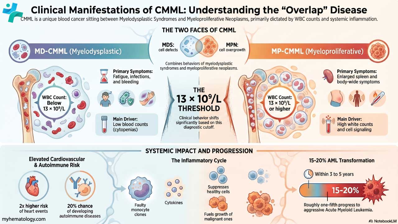
The headline diagnosis is chronic myelomonocytic leukemia (CMML), a disorder that blends features of myelodysplastic syndrome (poorly made, dysplastic blood cells) and myeloproliferative neoplasm (overproduction of myeloid cells). This is where current guidelines have shifted in a way older resources miss, so it deserves its own note below. A related childhood disorder, juvenile myelomonocytic leukemia, shares these overlapping features.
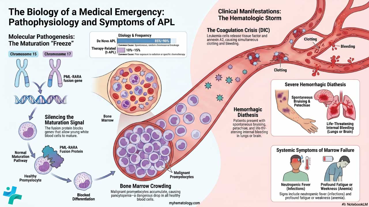
Acute myeloid leukemia (AML) with monocytic differentiation also features prominently; here the blood smear shows immature monocytes (monoblasts and promonocytes). Monocytosis can appear in other myeloproliferative neoplasms such as chronic myeloid leukemia and primary myelofibrosis, in Hodgkin and non-Hodgkin lymphoma (usually less strikingly), and occasionally with solid tumors through a paraneoplastic effect.
What Changed in 2022: The New CMML Threshold
For years, CMML required a monocyte count of at least 1.0 × 10⁹/L. In 2022, both the World Health Organization (5th edition) and the International Consensus Classification lowered that threshold to 0.5 × 10⁹/L [6,7]. Updated diagnostic criteria from 2022 reduce the monocyte threshold to 0.5 × 10⁹/L for CMML diagnosis, which will reclassify many cases previously diagnosed as MDS.
This change created a newly recognized group sometimes called oligomonocytic CMML, where the monocyte count sits in the 0.5–1.0 × 10⁹/L range with supporting evidence of clonality or dysplasia [11]. The practical message: a "borderline" persistent monocytosis is no longer automatically reassuring. Next-generation sequencing (NGS) is now a standard of care, as identifying specific somatic mutations (most commonly TET2, SRSF2, and ASXL1) provides definitive evidence of clonality and is crucial for diagnostic confirmation, risk stratification, and determining transplant eligibility [7,8].

Other Causes
A handful of other situations raise monocytes. After a splenectomy, monocytes that the spleen would normally remove stay in circulation. During recovery from bone marrow suppression (for example, after chemotherapy), monocytes often rebound first, causing a temporary rise. Severe physical stress, certain medications (including corticosteroids and the growth factor G-CSF), rare lipid storage diseases, and pregnancy can all contribute.
Clinical Presentation and Symptoms
This section is short for a reason. Monocytosis itself usually causes no symptoms. Whatever a patient feels comes from the underlying condition driving the count up. So the smart approach is to ask: what tends to cause monocytosis, and what would those conditions feel like?
When the cause is a chronic infection, expect features like low-grade persistent fever, fatigue, unexplained weight loss, and night sweats, sometimes with localizing clues such as a cough in tuberculosis. When the cause is an inflammatory or autoimmune disease, the picture leans toward fatigue, general malaise, joint pain or swelling, and skin rashes, plus symptoms specific to the organ involved.
When the cause is a blood cancer, the warning signs are more systemic. Classic "B symptoms" (unexplained fever, weight loss, and drenching night sweats) may appear, along with fatigue and pallor from anemia, easy bruising or bleeding from thrombocytopenia (low platelets), and recurrent infections from neutropenia (low neutrophils). An enlarged spleen or liver may also be present.
Persistent, high monocytosis (for example, above 1 × 10⁹/L for months) in someone who otherwise seems well should raise real suspicion for CMML, even before other symptoms appear [2]. Likewise, spotting immature monocytes on a smear is a strong visual clue pointing toward AML with monocytic differentiation.
Laboratory Investigations
A high monocyte count opens an investigation, and that investigation works best as a series of steps. The goal is to separate reactive causes from neoplastic ones, then pin down the specific diagnosis [3].
Step 1: History and Physical Examination
A careful history does much of the heavy lifting. Key questions cover the duration of the monocytosis (anything persisting beyond three months is more concerning), constitutional symptoms like fever and weight loss, infectious exposures such as travel or animal contact, autoimmune symptoms like joint pain or rashes, bleeding or recurrent infections that hint at marrow failure, and a medication and surgical history (including any past splenectomy). The physical exam looks for enlarged lymph nodes, an enlarged spleen or liver, skin lesions, and joint inflammation.
Step 2: Complete Blood Count and Blood Smear

The first lab step is a complete blood count with differential to confirm the AMC is genuinely elevated. Just as important is what the other cell lines are doing. Anemia, low or high platelets, and a high or low overall white count all add context. Monocytosis alongside low neutrophils or low counts across the board (pancytopenia) is especially suspicious for a marrow disorder.
The peripheral blood smear is then examined under the microscope. Reviewers look for dysplastic (abnormally formed) monocytes that suggest CMML, immature myeloid cells (blasts) that suggest AML, and abnormalities in red cells or other white cells that point to specific diagnoses.
Step 3: Targeted Testing
From here, testing follows suspicion. If infection is likely, that means cultures, serology, and imaging. If autoimmune disease is likely, that means autoantibody panels. If a blood cancer is on the table, the workup escalates to bone marrow biopsy, molecular and genetic testing (increasingly central, since genetic profiles now guide both diagnosis and prognosis [7]), and a specialized flow cytometry test.
That flow cytometry test deserves a mention because it is a genuinely modern tool, formally known as the monocyte partitioning assay. It sorts monocytes into three functionally distinct subsets based on the surface markers CD14 and CD16. In CMML, the classical monocyte population (CD14++/CD16-) expands substantially. Having greater than 94% classical monocytes is a highly sensitive and specific feature of CMML, and the 2022 WHO classification formally includes this abnormal partitioning as a supporting diagnostic criterion to reliably distinguish CMML from reactive monocytosis [6,9].
For mild, transient monocytosis with no worrying features, a simple repeat CBC in two to four weeks is often enough to confirm it has settled. If it persists or worsens, the fuller workup above is warranted.

Treatment and Management
The guiding rule is simple: you treat the cause, not the count. Monocytosis fades on its own once the underlying problem is brought under control. An infection calls for the right antibiotics; an autoimmune flare calls for appropriate immune-modulating therapy. Throughout, supportive care matters too, including fever control, nutritional support, and transfusions for severe anemia or low platelets.
The harder question is what happens when the cause is CMML, and here the original short answer of "treat the cause" needs filling in. There is no single easy cure, and management is tailored to the patient's age, fitness, and disease risk.
Many patients begin with supportive care or gentle treatments to control symptoms. Historically, the mainstay drug therapy has been the hypomethylating agents (HMAs) azacitidine and decitabine, though expectations must be realistic as only about 40% of patients achieve a transient response. Today, for high-risk CMML, combining an HMA with the BCL-2 inhibitor venetoclax is increasingly utilized to achieve better and deeper initial response rates than HMAs alone [8]. However, for suitable, usually younger or fitter patients, allogeneic stem cell transplantation remains the only potentially curative measure and the ultimate goal for durable remission [8].
Frequently Asked Questions (FAQs)
Is high monocytes the same as having cancer?
No. A high monocyte count is a sign, not a diagnosis. Most causes are non-cancerous, including chronic infections like tuberculosis, autoimmune diseases, and recovery after illness. Cancer (such as CMML) is one possibility a doctor checks for, especially when the count stays high for months without an obvious explanation.
What monocyte level is considered too high?
In adults, the normal absolute monocyte count is roughly 0.2–0.8 × 10⁹/L. A count above about 0.8–1.0 × 10⁹/L is generally called monocytosis. Importantly, blood cancer specialists now use a lower cutoff of 0.5 × 10⁹/L when specifically investigating chronic myelomonocytic leukemia, following the 2022 classification update.
How long does monocytosis need to last before doctors worry about leukemia?
Persistence is key. Monocytosis lasting longer than three months without a clear reactive cause raises more concern and usually prompts further testing, including a blood smear, molecular (genetic) testing, and sometimes a bone marrow biopsy.
How is the cause of high monocytes found?
Doctors start with a history, physical exam, and a complete blood count with a blood smear. From there, testing is targeted: cultures and imaging if infection is likely, autoantibody tests if autoimmune disease is suspected, and bone marrow biopsy plus genetic testing if a blood cancer is possible. A specialized flow cytometry "monocyte assay" can help identify CMML.
Can infections or vaccines raise monocytes?
Yes. Many chronic infections raise monocytes, and vaccines can cause a temporary, harmless rise as part of a normal immune response. These reactive increases typically settle once the trigger resolves.
How is high monocytes treated?
Treatment targets the underlying cause, not the count itself. An infection is treated with the right antibiotics; an autoimmune flare is treated with appropriate therapy. When the cause is CMML, options include supportive care, hypomethylating agents such as azacitidine, and allogeneic stem cell transplant, which is currently the only potential cure.
Glossary of Related Medical Terms
- Absolute monocyte count (AMC): The actual number of monocytes in a set volume of blood, found by multiplying the total white cell count by the monocyte percentage. It is the number that matters clinically.
- Monocyte: A type of white blood cell that swallows pathogens and debris, presents antigens to other immune cells, and matures into macrophages and dendritic cells in tissues.
- Macrophage: A long-lived "big eater" cell in tissues, derived from monocytes, that clears microbes and dead cells.
- Reactive (vs. neoplastic): "Reactive" means the count is rising in response to another problem like infection. "Neoplastic" means the cells themselves are part of a cancer.
- CMML (chronic myelomonocytic leukemia): A blood cancer with features of both myelodysplastic syndrome and myeloproliferative neoplasm, defined by long-standing high monocytes.
- Cytopenia: A low count of one or more blood cell types (red cells, white cells, or platelets).
- Dysplasia: Abnormal-looking, poorly formed blood cells, a clue to bone marrow disease.
- Hypomethylating agent (HMA): A drug class (azacitidine, decitabine) that changes how genes are switched on in cancer cells, used in CMML and related disorders.
- Allogeneic stem cell transplant: Replacing a patient's diseased bone marrow with stem cells from a donor; the only potential cure for CMML.
- Peripheral blood smear: A drop of blood spread on a slide and examined under a microscope to assess cell shape and maturity.
Disclaimer: This article is intended for educational and informational purposes only. It is not intended to be a substitute for informed professional medical advice, diagnosis, or treatment. While the information presented here is derived from credible medical sources and is believed to be accurate and up-to-date, it is not guaranteed to be complete or error-free. See additional information.
References
- Lynch, D. T., Hall, J., & Foucar, K. (2018). How I investigate monocytosis. International journal of laboratory hematology, 40(2), 107–114. https://doi.org/10.1111/ijlh.12776
- Christensen, M. E., Siersma, V., Kriegbaum, M., Lind, B. S., Samuelsson, J., Østgård, L. S. G., Grønbaek, K., & Andersen, C. L. (2023). Monocytosis in primary care and risk of haematological malignancies. European journal of haematology, 110(4), 362–370. https://doi.org/10.1111/ejh.13911
- Mangaonkar, A. A., Tande, A. J., & Bekele, D. I. (2021). Differential Diagnosis and Workup of Monocytosis: A Systematic Approach to a Common Hematologic Finding. Current hematologic malignancy reports, 16(3), 267–275. https://doi.org/10.1007/s11899-021-00618-4
- Dutta, P., & Nahrendorf, M. (2014). Regulation and consequences of monocytosis. Immunological reviews, 262(1), 167–178. https://doi.org/10.1111/imr.12219
- Gordon, S., & Taylor, P. R. (2005). Monocyte and macrophage heterogeneity. Nature reviews. Immunology, 5(12), 953–964. https://doi.org/10.1038/nri1733
- Khoury, J. D., Solary, E., Abla, O., Akkari, Y., Alaggio, R., Apperley, J. F., Bejar, R., Berti, E., Busque, L., Chan, J. K. C., Chen, W., Chen, X., Chng, W. J., Choi, J. K., Colmenero, I., Coupland, S. E., Cross, N. C. P., De Jong, D., Elghetany, M. T., Takahashi, E., … Hochhaus, A. (2022). The 5th edition of the World Health Organization Classification of Haematolymphoid Tumours: Myeloid and Histiocytic/Dendritic Neoplasms. Leukemia, 36(7), 1703–1719. https://doi.org/10.1038/s41375-022-01613-1
- Arber, D. A., Orazi, A., Hasserjian, R. P., Borowitz, M. J., Calvo, K. R., Kvasnicka, H. M., Wang, S. A., Bagg, A., Barbui, T., Branford, S., Bueso-Ramos, C. E., Cortes, J. E., Dal Cin, P., DiNardo, C. D., Dombret, H., Duncavage, E. J., Ebert, B. L., Estey, E. H., Facchetti, F., Foucar, K., … Tefferi, A. (2022). International Consensus Classification of Myeloid Neoplasms and Acute Leukemias: integrating morphologic, clinical, and genomic data. Blood, 140(11), 1200–1228. https://doi.org/10.1182/blood.2022015850
- Patnaik, M. M., & Tefferi, A. (2024). Chronic myelomonocytic leukemia: 2024 update on diagnosis, risk stratification and management. American journal of hematology, 99(6), 1142–1165. https://doi.org/10.1002/ajh.27271
- Liu, Y., Tariq, H., Fu, L., Gao, J., Jagtiani, T., Wolniak, K., Aqil, B., Ji, P., Chen, Y. H., & Chen, Q. C. (2025). Usefulness of Flow Cytometry Monocyte Partitioning in the Diagnosis of Chronic Myelomonocytic Leukemia in a Real-World Setting. Cancers, 17(7), 1229. https://doi.org/10.3390/cancers17071229