Key Takeaways
Severe malarial anemia is the most common life-threatening complication of malaria, defined by hemoglobin below 5 g/dL or hematocrit below 15% in children under 12, and below 7 g/dL (hematocrit below 20%) in adults [2].
- Mechanism ▾: The condition is driven by two processes happening at once: destruction of both infected and uninfected red blood cells, and suppression of red cell production in the bone marrow [3].
- Diagnosis ▾: Diagnosis combines clinical assessment, a thin and thick blood smear (still the gold standard), and a rapid diagnostic test, supported by a full blood count and reticulocyte count [6].
- Treatment ▾: Treatment requires intravenous or intramuscular artesunate for at least 24 hours, followed by a full three-day course of an artemisinin-based combination therapy; blood transfusion is indicated for severe anemia with decompensation [2].
- Prevention ▾: Prevention now includes two WHO-recommended vaccines (RTS,S/AS01 and R21/Matrix-M), insecticide-treated nets, and seasonal chemoprevention in high-transmission areas [1,8].
*Click ▾ for more information
Introduction
Severe malarial anemia is what a malaria infection looks like when it overwhelms the blood. It is the single most common way malaria kills young children, and for caregivers of a child returning from a malaria-endemic area, it is the complication that explains why fever is never something to wait out.
Malaria remains one of the largest infectious disease burdens in the world. The WHO World Malaria Report 2024 estimated 282 million cases and roughly 610,000 deaths in 2024, an increase of about 9 million cases compared with the previous year [1]. The African Region accounted for 95% of cases and 95% of deaths, and children under 5 made up about three-quarters of deaths in that region [1]. Severe malarial anemia is responsible for a substantial share of those pediatric deaths.
Five Plasmodium species infect humans: P. falciparum, P. vivax, P. malariae, P. ovale, and P. knowlesi. P. falciparum causes most severe disease, including most severe malarial anemia. P. vivax and P. knowlesi can also cause severe illness, and P. vivax has the distinctive ability to relapse from dormant liver forms months or years after the original infection [5].
Malaria Life Cycle
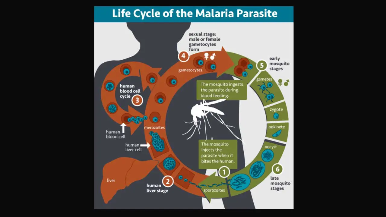
The malaria parasite alternates between a mosquito host and a human host. Understanding this cycle explains every part of malaria treatment and prevention.
Inside the mosquito (sporogonic cycle)
When a female Anopheles mosquito feeds on an infected person, it ingests sexual forms of the parasite called gametocytes. Inside the mosquito's midgut, these fuse and develop over 10–18 days into sporozoites, the infectious stage. The sporozoites migrate to the mosquito's salivary glands, ready to be injected into the next person bitten.
Inside the human (asexual cycle)
When the mosquito bites again, sporozoites enter the bloodstream and reach the liver within minutes. There, they invade liver cells and multiply for 7–14 days in a process called exo-erythrocytic schizogony, producing thousands of merozoites. P. vivax and P. ovale can also form dormant hypnozoites in the liver that reactivate weeks or years later [5].
When liver cells rupture, merozoites pour into the bloodstream and invade red blood cells. Inside the red cell, each parasite matures over about 48 hours, replicates, and bursts the cell to release a new wave of merozoites. This is the cycle that produces the classic fevers and the central driver of severe malarial anemia. Some merozoites instead develop into gametocytes, ready to be picked up by the next mosquito.
The repeated rupture of red cells, release of parasite molecules, and the immune response together produce the severe complications of malaria [5].
Defining Severe Malarial Anemia
The WHO definition for severe malarial anemia in a child under 12 is hemoglobin below 5 g/dL or hematocrit below 15%. In adults the threshold is hemoglobin below 7 g/dL or hematocrit below 20%. In pregnancy, hemoglobin below 7 g/dL with parasitemia is treated as severe [2,3].
These thresholds matter because severe malarial anemia rarely looks dramatic. Cerebral malaria draws attention with seizures and coma. Severe anemia can be quieter with pallor, breathlessness on feeding in an infant, fatigue, a fast heart rate and is sometimes missed until decompensation. In endemic areas it is the most common, and arguably the most lethal, complication of malaria.
Why the Anemia Becomes Severe
Two mechanisms work in parallel.
Destruction of red blood cells
Direct rupture of infected red cells is the obvious part, but it does not account for the depth of the anemia. For every infected cell destroyed, roughly eight uninfected red cells are also cleared from circulation [3]. Several factors make this happen: malaria infection changes the surface of healthy red cells (oxidation, reduced deformability, altered surface antigens), and the spleen's filtering activity rises sharply. Immune mechanisms for example complement deposition, autoantibodies, and immune complexes add to the destruction.
Reduced production of red blood cells
The bone marrow's response is blunted. In severe P. falciparum infection, the reticulocyte count is often inappropriately low, which means the marrow is not making enough new cells to replace those being lost [3]. Inflammatory mediators released early in infection, particularly tumor necrosis factor (TNF) and interferon-gamma (IFN-γ), make red cell precursors less responsive to erythropoietin (the hormone that drives red cell production). The result is dyserythropoiesis (defective red cell production) and a marrow that cannot keep up. Anemia can persist for weeks after the parasites are cleared, which is consistent with continuing destruction and slow marrow recovery [3].
The Role of Platelets
Thrombocytopenia (a low platelet count) is common in malaria and tends to be most marked in severe P. falciparum infection. The degree of thrombocytopenia correlates with parasite density and clinical outcome [4]. Platelet activation in malaria has two faces: activated platelets can bind to parasitized red cells and release platelet factor 4, which can kill the parasite inside; but the same activation can also contribute to microvascular blockage when infected red cells stick to vessel walls [4]. For the clinical reader, the practical point is simple: low platelets in a febrile patient with travel exposure should raise suspicion of malaria.
Symptoms
Early malaria looks like a flu. Within one to four weeks of a mosquito bite, patients typically develop fever and chills, sweating, headache, muscle aches, fatigue, nausea, and vomiting. The pattern can be intermittent, corresponding to the 48-hour replication cycle.
Severe disease announces itself with one or more of the following: severe anemia (pallor, breathlessness, fatigue), jaundice, seizures or impaired consciousness (cerebral malaria), kidney injury, respiratory distress, and dark urine.
The pattern of severe disease varies with age. Severe malarial anemia dominates in children under 2 years old. Older children are more likely to develop seizures and cerebral malaria. Adults face a broader spread of complications including acute kidney injury, pulmonary edema, and liver dysfunction.
How Severe Malarial Anemia Is Diagnosed
Diagnosis has two parts: confirming malaria and characterizing the anemia.
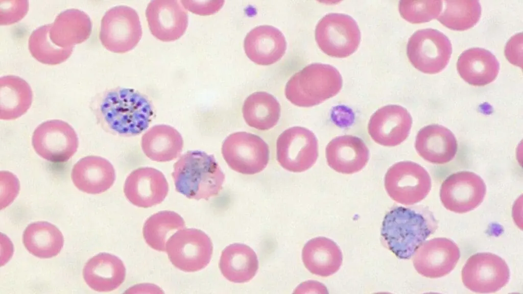
Confirming malaria. A Giemsa-stained thin and thick blood film remains the gold standard. Thick films are more sensitive for detecting parasites at low density; thin films allow species identification and parasitemia quantification [6]. Rapid diagnostic tests (RDTs) detect Plasmodium antigens such as HRP-2 (specific to P. falciparum) and can give an answer in 15–20 minutes, which is invaluable in resource-limited settings. RDTs are less sensitive at low parasite densities and cannot reliably quantify parasitemia [6]. PCR and loop-mediated isothermal amplification (LAMP) offer higher sensitivity and are useful for submicroscopic infections, mixed-species infections, and reference confirmation.
However, reliance on HRP-2 RDTs is increasingly threatened by Plasmodium falciparum parasites carrying pfhrp2 and pfhrp3 gene deletions, which cause false-negative diagnostic results. The global pooled prevalence of pfhrp2 deletions is estimated at over 21%, with high concentrations in South America and Africa, necessitating the use of alternative RDTs targeting Plasmodium lactate dehydrogenase (pLDH) or high-quality microscopy in affected regions [9].
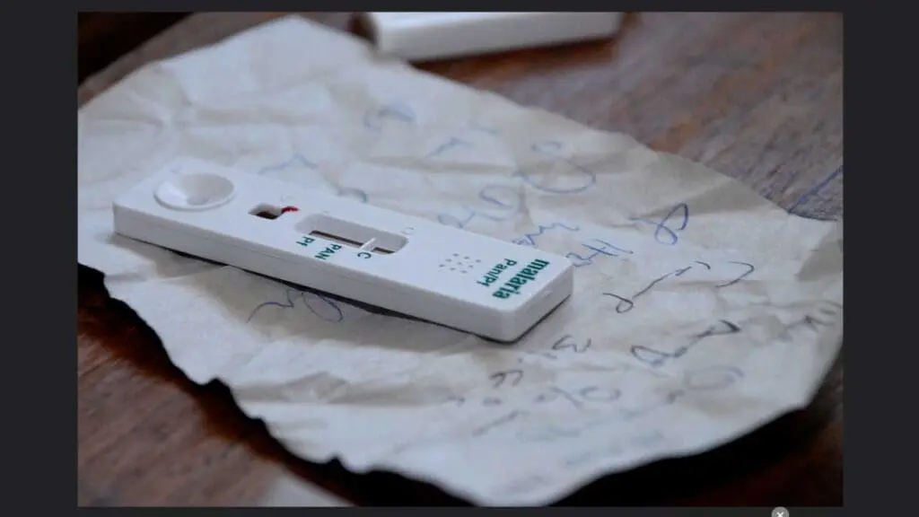
Characterizing the anemia. A full blood count establishes the severity. A reticulocyte count distinguishes a marrow that is responding from one that is not. In severe malarial anemia it is typically lower than the degree of anemia would predict. Markers of hemolysis (raised lactate dehydrogenase, raised indirect bilirubin, low haptoglobin) confirm red cell destruction. Renal and liver function tests, electrolytes, and glucose are essential because severe malaria affects multiple organs and hypoglycemia is common, especially in children and pregnant women.
Treatment
Treatment of severe malarial anemia has two simultaneous tracks: kill the parasite, and correct the anemia and its consequences.
Antimalarial therapy
WHO recommends intravenous or intramuscular artesunate as first-line treatment for severe malaria in adults, children, infants, and pregnant women in all trimesters [2]. Artesunate is given for at least 24 hours and continued until the patient can tolerate oral medication. Once oral therapy is possible, a full three-day course of an artemisinin-based combination therapy (ACT) completes treatment [2]. Children under 20 kg receive a higher artesunate dose per kilogram (3 mg/kg per dose) than larger children and adults (2.4 mg/kg per dose), to achieve equivalent drug exposure [2].
A newer one-step arginine-based formulation of injectable artesunate, prequalified by WHO in 2023, simplifies preparation and reduces dosing errors. Where IV/IM artesunate is not immediately available, a single dose of rectal artesunate can be given to children under 6 as pre-referral treatment before urgent transfer to a facility [2].
Where artesunate is unavailable, parenteral artemether or quinine are alternatives, though both are inferior. Quinine must be given as a slow controlled infusion because rapid administration is dangerous [2]. Artemisinin partial resistance has now been confirmed in P. falciparum in Eritrea, Rwanda, Uganda, and Tanzania, with suspicion in several other African countries; resistance to partner drugs such as piperaquine has emerged elsewhere [1,7]. This is why combination therapy and surveillance remain non-negotiable.
Correcting the anemia
WHO recommends blood transfusion for children with hemoglobin below 4 g/dL, or 4–6 g/dL with signs of decompensation (respiratory distress, impaired consciousness, hyperparasitemia, or shock) [2]. Transfusion in adults follows similar principles based on hemoglobin and clinical status. Exchange transfusion, once used as an adjunct in hyperparasitemia, is no longer recommended.
When transfusion is indicated, the volume must be managed carefully. A large randomized trial (TRACT) established that a strategy of giving 20 mL per kilogram of whole blood (or 10 mL per kilogram of packed red cells) remains standard, as higher volumes did not decrease mortality and must be carefully balanced against the risks of fluid overload and transfusion-transmitted infections [10].
Monitoring for post-artesunate delayed hemolysis
Patients who receive intravenous artesunate, particularly those with high parasite densities, can develop delayed hemolytic anemia 7–28 days after treatment. The mechanism involves synchronous removal of "pitted" red cells that survived parasite removal by the spleen. Follow-up FBC and hemolysis markers at one and four weeks after artesunate are recommended for any patient treated for severe malaria.
Supportive care
Severe malarial anemia rarely exists alone, and patients require highly restrictive fluid management because rapid intravenous fluid boluses have been shown to significantly increase 48-hour mortality in critically ill African children with impaired perfusion, often precipitating cardiovascular collapse or fatal pulmonary edema [11]. Therefore, cautious maintenance fluids should be used unless advanced, profound shock is present. The clinical team must also focus on the correction of hypoglycemia, treatment of seizures, monitoring of kidney function, and immediate empiric treatment of bacterial co-infections.
Prevention
Prevention of severe malarial anemia is prevention of malaria itself.
Vaccines. Two malaria vaccines are now WHO-recommended for use in children in regions of moderate to high P. falciparum transmission: RTS,S/AS01, recommended in 2021, and R21/Matrix-M, WHO-prequalified in 2023 [1,8]. By the end of 2024, 17 African countries had introduced a malaria vaccine into routine childhood immunization. Both vaccines are designed to reduce severe disease and death rather than to interrupt transmission [1].
Vector control. Insecticide-treated nets remain the foundation. Pyrethroid-PBO nets and dual active-ingredient nets are recommended where pyrethroid resistance is documented [2]. Indoor residual spraying complements bed nets.
Chemoprevention. Seasonal malaria chemoprevention for children in the Sahel, intermittent preventive treatment in pregnancy, and perennial malaria chemoprevention for infants in high-transmission areas all reduce infection and severe anemia in target groups [2].
For travelers. Chemoprophylaxis appropriate for the destination, started before travel and continued for the required duration after return, is essential. Bite avoidance using repellents, long sleeves at dusk and dawn, and bed nets is part of the same package.
Frequently Asked Questions (FAQs)
What is severe malarial anemia?
Severe malarial anemia is a life-threatening drop in red blood cells caused by malaria infection. The World Health Organization defines it as a hemoglobin level below 5 g/dL or a hematocrit below 15% in children under 12, and below 7 g/dL (hematocrit below 20%) in adults. It is the most common severe complication of malaria in young African children and is caused by both destruction of red blood cells and reduced production in the bone marrow.
Which malaria parasite causes severe malarial anemia?
Plasmodium falciparum is responsible for the majority of severe malarial anemia cases worldwide, especially in sub-Saharan Africa. Plasmodium vivax can also cause severe anemia, particularly with repeated or untreated infections, and Plasmodium knowlesi can cause severe disease in Southeast Asia.
How is severe malarial anemia treated?
Treatment has two parts. First, the parasite is cleared with intravenous or intramuscular artesunate for at least 24 hours, followed by a full three-day course of an oral artemisinin-based combination therapy once the patient can swallow. Second, the anemia itself is corrected: WHO recommends blood transfusion in children with hemoglobin below 4 g/dL, or 4–6 g/dL with signs such as breathlessness or impaired consciousness. Patients also need monitoring for delayed hemolysis after artesunate.
Is there a vaccine for malaria?
Yes. Two malaria vaccines are WHO-recommended: RTS,S/AS01 (recommended in 2021) and R21/Matrix-M (prequalified in 2023). Both target P. falciparum and are designed to reduce severe disease and death in young children. By the end of 2024, 17 African countries had introduced a malaria vaccine into their routine childhood immunization schedule.
Why does malaria cause such severe anemia in children?
Children, especially those under 5 in malaria-endemic areas, often face repeated infections before they develop partial immunity. Each infection destroys both infected and uninfected red blood cells, while inflammation suppresses red cell production in the bone marrow. Co-existing iron deficiency, undernutrition, and infections such as hookworm make the anemia worse. This combination explains why severe malarial anemia is the leading cause of malaria death in young African children.
How can severe malarial anemia be prevented?
Prevention works on two fronts: stopping mosquito bites and treating early infections before they progress. Insecticide-treated bed nets, indoor residual spraying, and seasonal malaria chemoprevention for children in high-transmission areas reduce infections. Malaria vaccines reduce severe disease in young children. Travelers to endemic areas should take chemoprophylaxis recommended for their destination. Prompt diagnosis with a blood smear or rapid diagnostic test, and prompt treatment with an ACT, prevents progression to severe anemia.
Glossary of Related Medical Terms
- Anemia — A reduction in red blood cells or hemoglobin below the normal range for age and sex, leading to reduced oxygen delivery to tissues.
- Anopheles mosquito — The genus of mosquito whose female members transmit Plasmodium parasites to humans.
- Artemisinin-based combination therapy (ACT) — A treatment that pairs a fast-acting artemisinin drug with a longer-acting partner drug to clear malaria and slow resistance.
- Cytoadhesion — The sticking of infected red blood cells to blood vessel walls, central to severe malaria pathology.
- Erythropoiesis — The production of red blood cells, mainly in the bone marrow.
- Erythropoietin (EPO) — The hormone, made mostly by the kidneys, that drives red blood cell production.
- Gametocyte — The sexual stage of the malaria parasite, picked up by mosquitoes from infected people.
- Hemoglobin — The iron-containing protein in red blood cells that carries oxygen.
- Hypnozoite — A dormant liver-stage form of Plasmodium vivax and P. ovale that can reactivate weeks to years later.
- Merozoite — The asexual stage released when infected liver cells or red blood cells rupture; it invades new red blood cells.
- Parasitemia — The percentage or count of red blood cells infected with parasites.
- Reticulocyte — A young red blood cell; a low reticulocyte count in anemia suggests the marrow is not responding.
- Sequestration — The build-up of infected red blood cells in small blood vessels of organs such as the brain.
- Sporozoite — The infectious stage of Plasmodium injected by a mosquito into human skin.
- Thrombocytopenia — A low platelet count, common in malaria and a marker of disease severity.
Disclaimer: This article is intended for educational and informational purposes only. It is not intended to be a substitute for informed professional medical advice, diagnosis, or treatment. While the information presented here is derived from credible medical sources and is believed to be accurate and up-to-date, it is not guaranteed to be complete or error-free. See additional information.
References
- World Health Organization. (2024). World malaria report 2024. https://www.who.int/teams/global-malaria-programme/reports/world-malaria-report-2024
- World Health Organization. (2024). WHO guidelines for malaria, 30 November 2024. https://www.who.int/publications/i/item/guidelines-for-malaria
- White N. J. (2018). Anaemia and malaria. Malaria journal, 17(1), 371. https://doi.org/10.1186/s12936-018-2509-9
- O'Sullivan, J. M., & O'Donnell, J. S. (2018). Platelets in malaria pathogenesis. Blood, 132(12), 1222–1224. https://doi.org/10.1182/blood-2018-08-865618
- Wassmer, S. C., Taylor, T. E., Rathod, P. K., Mishra, S. K., Mohanty, S., Arevalo-Herrera, M., Duraisingh, M. T., & Smith, J. D. (2015). Investigating the Pathogenesis of Severe Malaria: A Multidisciplinary and Cross-Geographical Approach. The American journal of tropical medicine and hygiene, 93(3 Suppl), 42–56. https://doi.org/10.4269/ajtmh.14-0841
- Fitri, L. E., Widaningrum, T., Endharti, A. T., Prabowo, M. H., Winaris, N., & Nugraha, R. Y. B. (2022). Malaria diagnostic update: From conventional to advanced method. Journal of clinical laboratory analysis, 36(4), e24314. https://doi.org/10.1002/jcla.24314
- Ippolito, M. M., Moser, K. A., Kabuya, J. B., Cunningham, C., & Juliano, J. J. (2021). Antimalarial Drug Resistance and Implications for the WHO Global Technical Strategy. Current epidemiology reports, 8(2), 46–62. https://doi.org/10.1007/s40471-021-00266-5
- Draper, S. J., Sack, B. K., King, C. R., Nielsen, C. M., Rayner, J. C., Higgins, M. K., Long, C. A., & Seder, R. A. (2018). Malaria Vaccines: Recent Advances and New Horizons. Cell host & microbe, 24(1), 43–56. https://doi.org/10.1016/j.chom.2018.06.008
- Jejaw Zeleke, A., Hailu, A., Bayih, A. G., Kefale, M., Amare, A. T., Tegegne, Y., & Aemero, M. (2022). Plasmodium falciparum histidine-rich protein 2 and 3 genes deletion in global settings (2010-2021): a systematic review and meta-analysis. Malaria journal, 21(1), 26. https://doi.org/10.1186/s12936-022-04051-7
- Maitland, K., Kiguli, S., Opoka, R. O., Engoru, C., Olupot-Olupot, P., Akech, S. O., Nyeko, R., Mtove, G., Reyburn, H., Lang, T., Brent, B., Evans, J. A., Tibenderana, J. K., Crawley, J., Russell, E. C., Levin, M., Babiker, A. G., Gibb, D. M., & FEAST Trial Group (2011). Mortality after fluid bolus in African children with severe infection. The New England journal of medicine, 364(26), 2483–2495. https://doi.org/10.1056/NEJMoa1101549
- Maitland, K., Olupot-Olupot, P., Kiguli, S., Chagaluka, G., Alaroker, F., Opoka, R. O., Mpoya, A., Engoru, C., Nteziyaremye, J., Mallewa, M., Kennedy, N., Nakuya, M., Namayanja, C., Kayaga, J., Uyoga, S., Kyeyune Byabazaire, D., M'baya, B., Wabwire, B., Frost, G., Bates, I., … TRACT Group (2019). Transfusion Volume for Children with Severe Anemia in Africa. The New England journal of medicine, 381(5), 420–431. https://doi.org/10.1056/NEJMoa1900100