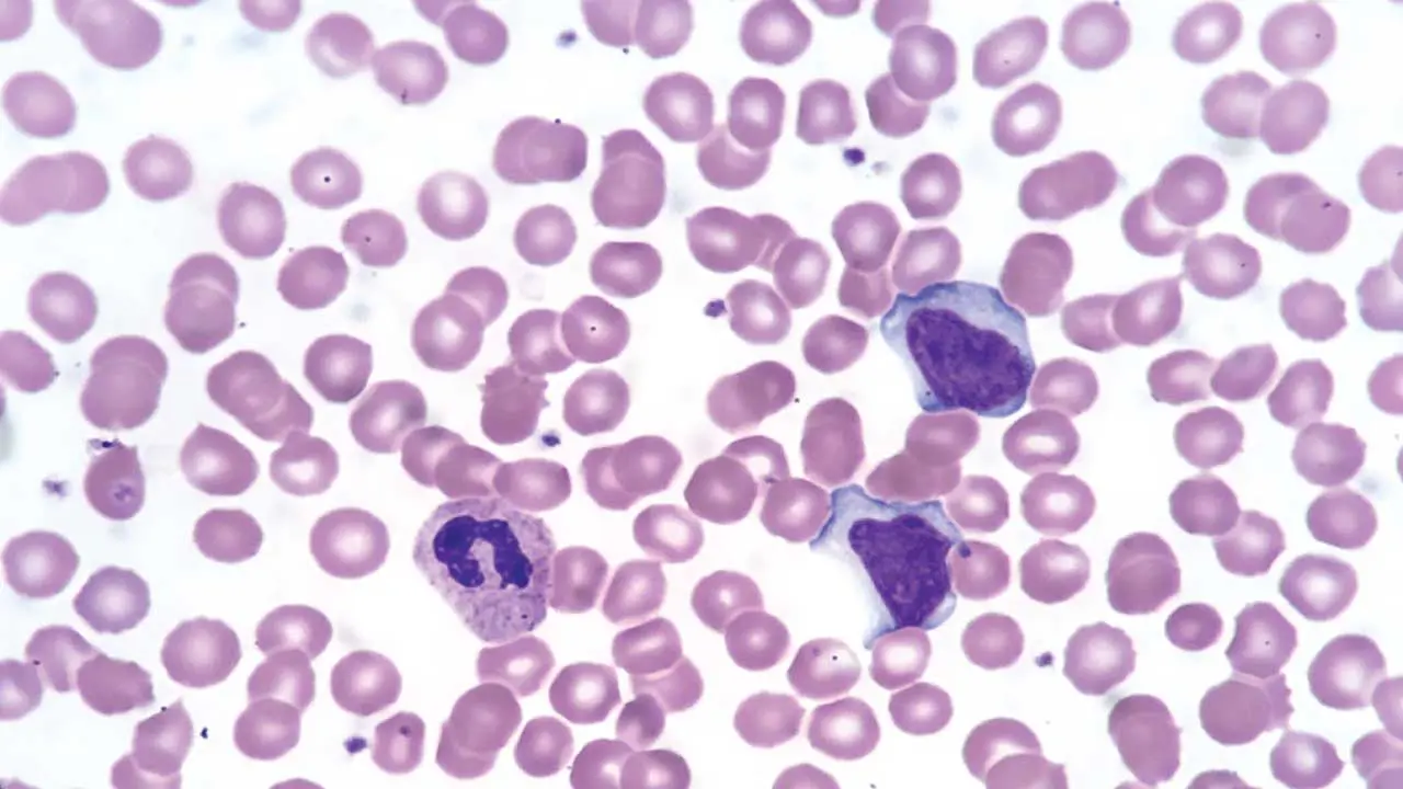
Key Takeaways
A bone marrow aspirate is a liquid sample of marrow cells used to assess cell morphology, while a trephine biopsy is a solid core of bone and marrow used to assess overall architecture and cellularity. The two are usually performed together [1,2].
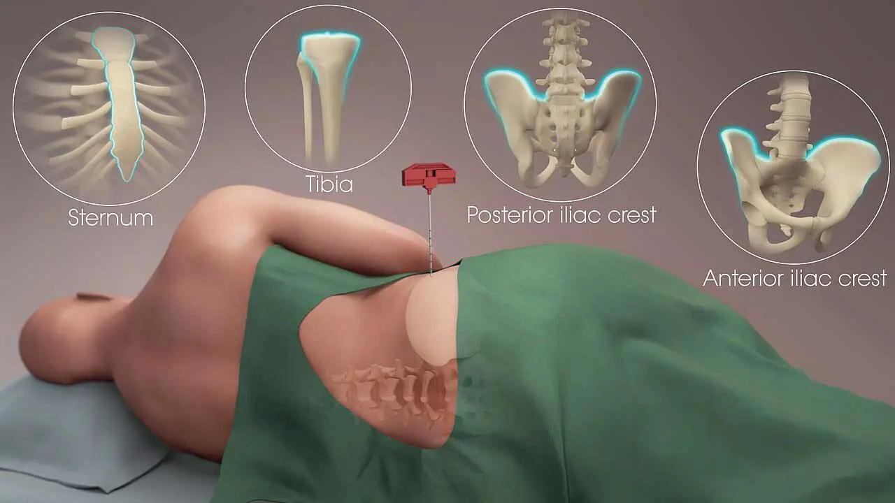
- Site ▾: The posterior superior iliac spine is the preferred site in adults and older children. The sternum can be used for aspiration only, never for trephine biopsy, because of the risk to underlying structures [1].
- Indications ▾: Common indications include unexplained cytopenias, suspected leukemia or lymphoma, staging of known malignancy, unexplained anemia, and investigation of fever of unknown origin [3,4].
- Interpretation ▾: A well-prepared bone marrow aspirate smear should be assessed for cellularity, the myeloid-to-erythroid (M:E) ratio, blast percentage, and megakaryocyte number, typically over a 500-cell differential count [4,5].
- Common pitfalls ▾: A "dry tap," or failure to obtain aspirate material, is most often caused by marrow fibrosis, densely packed marrow, or technical error, and it always warrants a trephine biopsy [1,5].
- Complications ▾: Major complications, such as significant bleeding or infection, are rare when the procedure is performed correctly with attention to site selection and post-procedure pressure.
*Click ▾ for more information
Why Bone Marrow Examination Matters
Blood tests can tell you what is wrong with a patient's blood counts. A bone marrow aspirate and trephine biopsy often tell you why. Blood cells are produced in the marrow before they are ever released into circulation, so when the peripheral blood picture doesn't add up, the marrow is where the answer usually lies.
Together, the aspirate and biopsy give complementary information. The bone marrow aspirate provides individual cells spread thinly on a slide, ideal for looking closely at cell shape and detail. The trephine biopsy provides an intact core of bone and marrow tissue, ideal for judging overall cellularity, architecture, and patterns of infiltration that loose cells cannot show [1,2]. Neither sample fully replaces the other, which is why both are usually taken in the same sitting.
| Characteristic | Aspiration | Trephine biopsy |
|---|---|---|
| Sample type | Liquid suspension of marrow cells and sinusoidal blood. | Solid core of intact bone and marrow tissue. |
| Primary clinical use | Detailed cell morphology and differential counts. | Overall marrow architecture and spatial distribution. |
| What it assesses best | Individual cell shape, blast percentage, and M:E ratio. | Overall cellularity, focal lesions, and fibrosis. |
| Preparation method | Spread thinly onto glass slides (smears) at the bedside. | Fixed in formalin, decalcified, and embedded in paraffin. |
| Turnaround time | Rapid 2–6 hours for initial smear review. | Slower 2–3 days due to decalcification processing. |
| Ancillary testing | Ideal for flow cytometry, cytogenetics, and molecular testing. | Used for immunohistochemistry and reticulin stains. |
| Impact of "dry tap" | Fails Cannot be used for morphology. | Preferred Becomes the primary diagnostic tool; reliably yields tissue. |
Indications for Bone Marrow Examination
A bone marrow aspirate and trephine biopsy may be requested for:
- Unexplained anemia, leukopenia, thrombocytopenia, or pancytopenia not explained by peripheral blood findings.
- Suspected acute or chronic leukemia, based on blood counts or blood film abnormalities.
- Staging and monitoring of lymphoma, multiple myeloma, or other known hematological malignancies.
- Investigation of unexplained fever, especially in immunocompromised patients (fever of unknown origin).
- Evaluation of suspected myelodysplastic syndrome (MDS) or myeloproliferative neoplasms.
- Assessment of iron stores when serum markers are unclear.
- Investigation of suspected marrow infiltration by metastatic cancer, granulomatous disease, or storage disorders [3,4].
While traditional morphology remains central, Next-Generation Sequencing (NGS) and Fluorescence in situ Hybridization (FISH) are now standard diagnostic pillars. Modern hematopathology relies heavily on high-throughput NGS for accurate diagnosis, prognostication, and targeted therapy selection, effectively complementing and expanding upon standard morphological workups [6,7].
Materials and Equipment
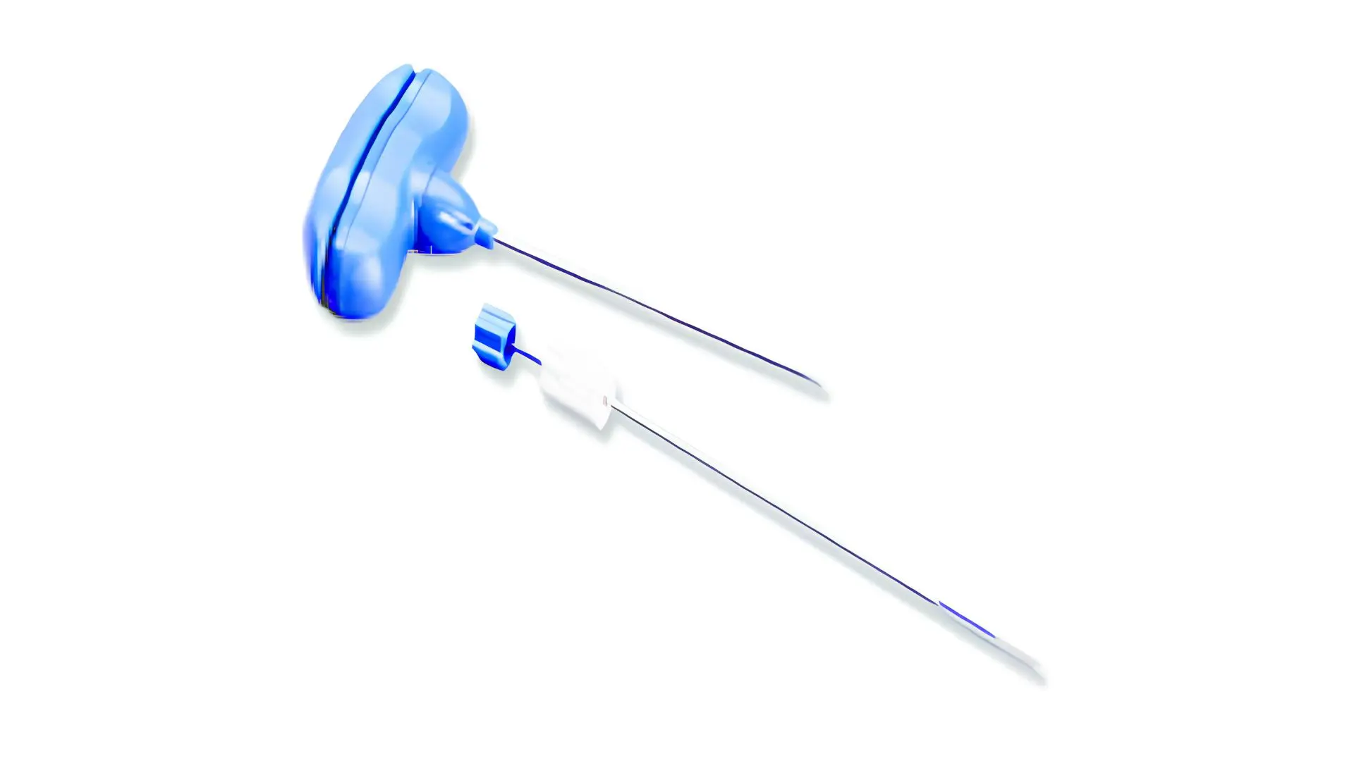
- Bone marrow aspiration and biopsy needle set (commonly a Jamshidi-type needle)
- Local anesthetic (lidocaine), syringes, and needles for infiltration
- Sterile drapes, gloves, and antiseptic skin preparation
- Glass slides for aspirate smears and particle crush preparations
- EDTA tubes for flow cytometry, cytogenetics, and molecular studies as required
- Culture media, if infection is suspected
- Fixative for the trephine biopsy core (typically formalin)
- Sterile dressing for the site after the procedure
Protocol
- Confirm patient identity, indication, and consent. Review coagulation status if clinically relevant.
- Position the patient. For a posterior iliac crest approach, the patient lies prone or on their side with the hip slightly flexed.
- Prepare the skin with antiseptic solution and apply a sterile drape.
- Infiltrate local anesthetic into the skin, subcutaneous tissue, and periosteum overlying the posterior superior iliac spine.
- Advance the aspiration needle through the cortex into the marrow cavity using a steady rotating motion.
- Once correctly positioned, remove the inner stylet and attach a syringe. Apply firm, brief suction to draw a small volume of marrow (ideally under 0.2 to 0.5 mL for morphology, to limit dilution with peripheral blood).
- Prepare smears immediately at the bedside: several thin smears, plus particle crush (squash) preparations, which best preserve marrow architecture at the cellular level.
- If additional marrow is needed for flow cytometry, cytogenetics, molecular testing, or culture, redirect the needle slightly and aspirate further small volumes into the appropriate tubes.
- For the trephine biopsy, reinsert the stylet or use a dedicated biopsy needle. Advance through a separate cortical entry point (or redirect from the aspiration site) with a controlled rotating motion to collect an intact core, ideally 1.5 to 2 cm in length.
- Withdraw the needle, apply firm pressure to the site for several minutes, then apply a sterile dressing.
- Observe the patient briefly for bleeding or discomfort before discharge from the procedure area [1,2].
WHO Criteria for Biopsy Core Adequacy
Advancing the needle with a controlled rotating motion secures a solid core. The World Health Organization (WHO) and diagnostic guidelines emphasize that biopsies must be non-tangential and measure at least 1.5 cm in length to be considered diagnostically adequate, ideally capturing multiple partially preserved intertrabecular areas [8].
Site Selection
| Site | Use | Notes |
|---|---|---|
| Posterior superior iliac spine | Aspirate and trephine biopsy | Preferred site in adults and children able to tolerate positioning. |
| Anterior iliac crest | Aspirate and trephine biopsy | Used when posterior access is difficult, or in obese patients. |
| Sternum | Aspirate only | Never used for trephine biopsy due to the risk to underlying mediastinal structures. |
| Tibia | Aspirate and trephine biopsy | Used mainly in infants under 1 year of age. |
Interpretation: Basic Morphological Assessment
Interpreting the sample follows a logical sequence, moving from overall appearance down to individual cells.
Step 1: Assess adequacy. A good aspirate smear should contain visible marrow particles (fragments of stroma and cells) and a thin trailing edge, similar in principle to a peripheral blood smear. A trephine biopsy core should be at least 1.5 cm long for reliable assessment, since shorter cores can underestimate cellularity or miss focal disease.
Step 2: Assess overall cellularity. On the trephine biopsy, cellularity is estimated as the proportion of marrow space occupied by hematopoietic cells versus fat. A rough rule of thumb is that normal cellularity approximates 100 minus the patient's age in percent, though this varies with sampling site and technique [5].
Calculate Cellularity (Trephine)
Cellularity is the ratio of blood-forming cells to fat spaces. A standard, widely used baseline formula for normal adult cellularity is:
100−age = Expected Cellularity %
(e.g., A 60-year-old should have roughly 40% cellularity). However, recent large-scale modeling demonstrates that the age-dependent decline in adults is actually slower than this classic formula suggests, dropping at approximately 3% per decade. This means cellularity often remains much more stable into extreme old age than previously assumed [9].
Step 3: Assess the myeloid-to-erythroid (M:E) ratio. On the aspirate, granulocyte precursors are compared to red cell precursors. The normal M:E ratio is approximately 2:1 to 4:1. A high ratio suggests granulocytic hyperplasia or erythroid suppression; a low ratio suggests the opposite [4,5].
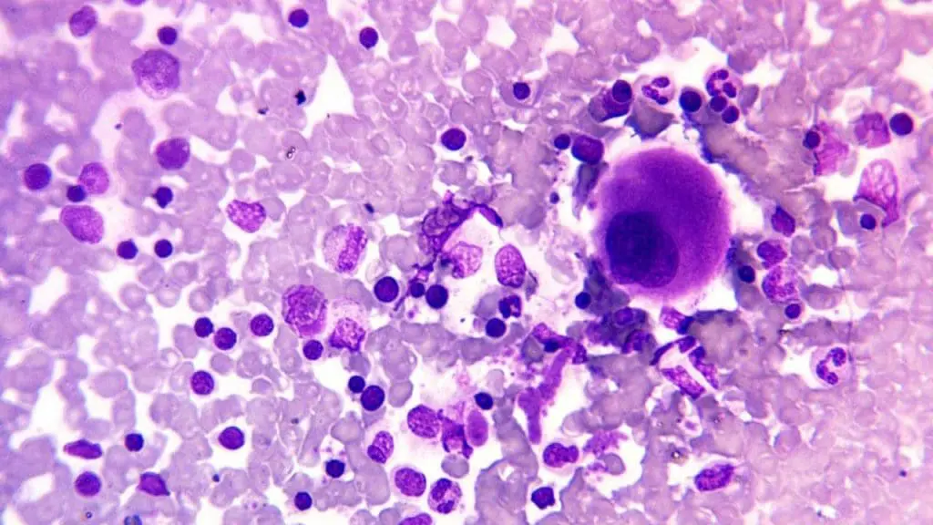
Step 4: Perform a differential count. A count of 500 nucleated cells on the aspirate smear characterizes the proportion of blasts, promyelocytes, myelocytes, band forms, segmented neutrophils, erythroid precursors, lymphocytes, plasma cells, and monocytes. Blast percentage is a key diagnostic threshold in classifying acute leukemia.
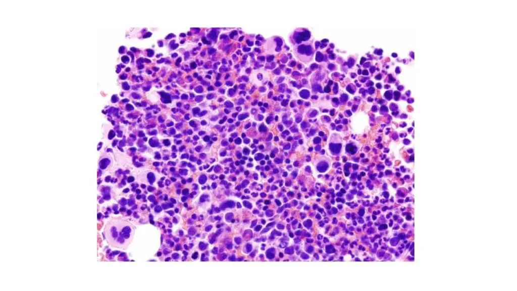
Step 5: Assess megakaryocytes. Megakaryocyte number and morphology (size, lobulation, clustering) are best judged on both the aspirate particles and the trephine biopsy, since megakaryocytes are often under-represented on thin smear areas.
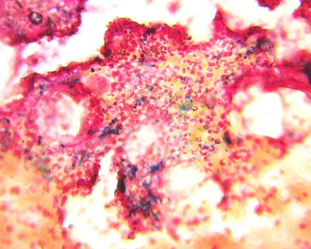
Step 6: Assess iron stores. A Perls' Prussian blue stain on the aspirate smear or particle preparation identifies stainable iron in macrophages, useful in the workup of anemia.
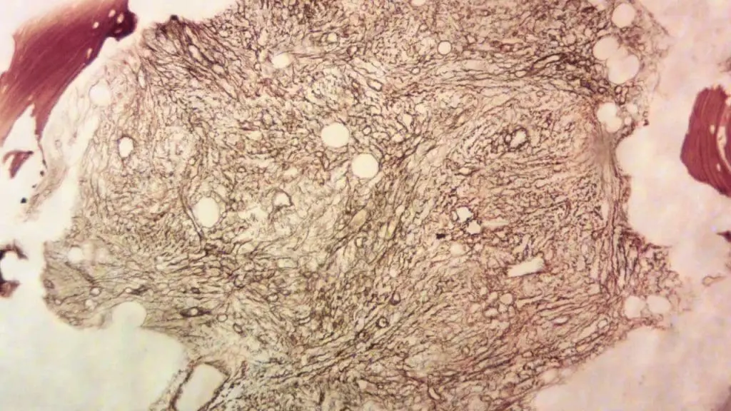
Step 7: Note architecture and infiltration patterns on the trephine biopsy. Patterns such as diffuse, focal, interstitial, or paratrabecular infiltration help distinguish reactive changes from lymphoma, myeloma, or metastatic disease. A reticulin stain assesses marrow fibrosis, relevant in myelofibrosis and some myeloproliferative neoplasms [1,5].
Common Pitfalls and Technical Considerations
| Problem | Likely Cause | Corrective Action |
|---|---|---|
| Dry tap (no aspirate obtained) | Marrow fibrosis, densely packed or hypercellular marrow, or needle malposition. | Proceed to trephine biopsy; consider repeat attempt at a different site if aspirate is essential. |
| Blood-diluted aspirate | Excessive suction volume or prolonged aspiration. | Limit initial aspirate to under 0.5 mL; use separate small aliquots for each additional test. |
| Crushed or distorted cells on smear | Excessive pressure while spreading, or delayed smear preparation. | Prepare smears immediately at the bedside with a light touch. |
| Fragmented or short trephine core | Needle redirected during advancement, or brittle bone. | Use a steady rotating motion without lateral force; consider a repeat pass. |
| Poor staining of aspirate smear | Smear too thick, or delayed fixation. | Prepare thin smears promptly and fix without delay. |
| Excessive bleeding at the site | Underlying coagulopathy, or inadequate post-procedure pressure. | Review coagulation status beforehand; apply firm pressure for several minutes after the procedure. |
Complications and Safety Considerations
Bone marrow aspiration and trephine biopsy are generally safe procedures with a low complication rate. The most common issues are localized pain and minor bruising at the site. Bleeding requiring intervention and infection are rare, but the risk increases in patients with significant thrombocytopenia, coagulopathy, or immunosuppression. Relative contraindications include severe, uncorrected bleeding disorders and infection overlying the intended site. Careful site selection, sterile technique, and adequate post-procedure pressure minimize risk in almost all patients [1,2].
Frequently Asked Questions
What is the difference between a bone marrow aspirate and a trephine biopsy?
A bone marrow aspirate is a liquid sample of marrow cells drawn out with a syringe, used to study individual cell shape and detail. A trephine biopsy is a solid core of bone and marrow tissue, used to assess overall cellularity and architecture. They are usually performed together at the same visit because each provides information the other cannot.
Where is a bone marrow aspirate usually taken from?
The posterior superior iliac spine is the preferred site in adults and older children. The sternum can be used for aspiration only, never for a trephine biopsy, because of the risk to the structures beneath it. In infants under one year, the tibia is sometimes used instead.
Why does a "dry tap" happen, and what does it mean?
A dry tap means no marrow fluid could be drawn despite correct needle placement. It is most often caused by marrow fibrosis or a densely packed marrow, both of which can pack the cells too tightly to aspirate. When this happens, the trephine biopsy becomes especially important, since it can still yield diagnostic tissue.
Is a bone marrow aspirate painful?
Local anesthetic numbs the skin and periosteum before the procedure, so most patients feel pressure rather than sharp pain. The moment of aspiration itself, when suction is applied, can cause a brief, sharp cramping sensation that typically lasts only a few seconds.
What does the myeloid-to-erythroid ratio tell you?
The M:E ratio compares the number of developing white blood cells to developing red blood cells in the marrow. A normal ratio is roughly 2:1 to 4:1. A ratio higher than normal suggests overproduction of white cells or suppression of red cell production, while a lower ratio suggests the reverse.
How long does it take to get results?
Aspirate smears can often be reviewed within a day or two. The trephine biopsy requires decalcification and processing before sectioning, which typically takes several days longer, so a complete report combining both samples usually takes about a week.
Glossary of Related Medical Terms
- Aspirate: A liquid sample of bone marrow cells drawn out through a needle and syringe.
- Cellularity: The proportion of the marrow space occupied by blood-forming cells, as opposed to fat.
- Dry tap: A failed attempt to draw marrow fluid, often due to fibrosis or densely packed marrow.
- M:E ratio (myeloid-to-erythroid ratio): The ratio of developing white blood cells to developing red blood cells in the marrow, normally about 2:1 to 4:1.
- Megakaryocyte: The large marrow cell responsible for producing platelets.
- Particle crush preparation: A smear made by crushing a marrow fragment between two slides, useful for assessing marrow architecture at the cellular level.
- Perls' stain: A stain used to detect stainable iron stores in marrow macrophages.
- Reticulin stain: A stain used on the trephine biopsy to assess the degree of marrow fibrosis.
- Trephine biopsy: A solid core of bone and marrow tissue removed for histological examination.
Disclaimer: This protocol is for educational purposes only. Local laboratory standard operating procedures take precedence. It is not intended to be a substitute for informed professional medical advice, diagnosis, or treatment. Always consult a qualified healthcare professional for clinical decision-making. While the information presented here is derived from credible medical sources and is believed to be accurate and up-to-date, it is not guaranteed to be complete or error-free. See additional information.
References
- Bain B. J. (2001). Bone marrow trephine biopsy. Journal of clinical pathology, 54(10), 737–742. https://doi.org/10.1136/jcp.54.10.737
- Bain B. J. (2001). Bone marrow aspiration. Journal of clinical pathology, 54(9), 657–663. https://doi.org/10.1136/jcp.54.9.657
- Malempati, S., Joshi, S., Lai, S., Braner, D. A., & Tegtmeyer, K. (2009). Bone marrow aspiration and biopsy. New England Journal of Medicine, 361(15), e28. https://www.nejm.org/doi/full/10.1056/NEJMvcm0804634
- Riley, R. S., Hogan, T. F., Pavot, D. R., Forysthe, R., Massey, D., Smith, E., Wright, L., Jr, & Ben-Ezra, J. M. (2004). A pathologist's perspective on bone marrow aspiration and biopsy: I. Performing a bone marrow examination. Journal of clinical laboratory analysis, 18(2), 70–90. https://doi.org/10.1002/jcla.20008
- Riley, R. S., Williams, D., Ross, M., Zhao, S., Chesney, A., Clark, B. D., & Ben-Ezra, J. M. (2009). Bone marrow aspirate and biopsy: a pathologist's perspective. II. interpretation of the bone marrow aspirate and biopsy. Journal of clinical laboratory analysis, 23(5), 259–307. https://doi.org/10.1002/jcla.20305
- Haferlach T. (2021). The time has come for next-generation sequencing in routine diagnostic workup in hematology. Haematologica, 106(3), 659–661. https://doi.org/10.3324/haematol.2020.270504
- Hasemeier, B., Geffers, R., Bartels, S., Schlegelberger, B., Kreipe, H., & Lehmann, U. (2013). Archival bone marrow trephines are suitable for high-throughput mutation analysis using next generation sequencing technology. Haematologica, 98(9), e115–e116. https://doi.org/10.3324/haematol.2013.091652
- Tripodo, C., Di Bernardo, A., Ternullo, M. P., Guarnotta, C., Porcasi, R., Ingrao, S., Gianelli, U., Boveri, E., Iannitto, E., Franco, G., & Florena, A. M. (2009). CD146(+) bone marrow osteoprogenitors increase in the advanced stages of primary myelofibrosis. Haematologica, 94(1), 127–130. https://doi.org/10.3324/haematol.13598
- Wong, J., Jackson, R., Chen, L., Song, J., Pillai, R., Afkhami, M., Danilova, O., Aoun, P., Gaal, K. K., & Kim, Y. (2024). Determination of age-dependent bone marrow normocellularity. American journal of clinical pathology, 161(2), 170–176. https://doi.org/10.1093/ajcp/aqad129