Procedure At-A-Glance
The Wright stain is a Romanowsky-type stain used to differentiate blood cell types and detect abnormalities on a peripheral blood smear.
Flood Slide Protocol
- Air-dry a well-made smear. Optionally fix in methanol.
- Flood the slide with undiluted Wright stain. Wait 1–3 minutes.
- Add an equal volume of buffer (pH 6.5–6.8). A green metallic sheen confirms correct mixing.
- Stain for 3–5 minutes.
- Rinse with buffer or distilled water until the thin edges turn pinkish-red.
- Air-dry the slide.
- Examine under oil immersion.
Dip Slide Protocol with Coplin Jars
- Fix the smear in methanol for 15–30 seconds.
- Dip in Wright stain for 2–3 minutes.
- Transfer to buffer (pH 6.8) for 1–4 minutes. Do not agitate.
- Rinse in buffer or distilled water until edges are pinkish-red.
- Air-dry and examine.
Introduction
Automated analyzers can count blood cells in seconds, but they cannot always identify what kind of abnormal cell they have flagged. The Wright stain bridges that gap. By coloring different parts of a cell in contrasting shades, it lets a trained eye classify cell types, recognize abnormal shapes, and spot blood-borne parasites.
The Wright stain is used mainly in two settings:
- Hematology, to evaluate red cells, white cells, and platelets when a blood count is abnormal, when anemia or hemolysis is suspected, or when leukemia is on the differential.
- Bone marrow assessment, though the closely related Wright-Giemsa stain is often preferred there for its richer nuclear detail.
Once stained, a smear can reveal immature cells, abnormal nuclear shapes, and unusual inclusions that point toward conditions such as leukemia, lymphoma, or anemia [3].
If you are new to blood films, start with our companion article on Preparation of Peripheral Blood Smears. A poorly made smear cannot be rescued by even the best stain.
When Is a Peripheral Blood Smear Indicated?
Knowing when to stain a smear matters as much as knowing how. A smear is typically requested when:
- An automated count flags abnormal cells, platelets, or red cell indices.
- A patient has unexplained anemia, jaundice, or suspected hemolysis.
- Blasts or atypical lymphocytes are reported by an analyzer.
- A blood-borne parasite is suspected.
- Diagnostic shapes such as schistocytes, sickle cells, or teardrop cells need confirmation.
Principle of the Wright Stain
The Wright stain belongs to the Romanowsky family of blood stains. It is a polychromatic stain, meaning it contains more than one dye. The two active components are methylene blue (and its oxidized azure derivatives), a basic blue dye, and Eosin Y, an acidic orange-red dye [5]. Together they exploit one simple rule of chemistry: opposite charges attract.
- Methylene blue and azures are positively charged (basic). They bind negatively charged structures, especially the DNA in nuclei and the RNA in cytoplasm. These appear blue, violet, and purple.
- Eosin Y is negatively charged (acidic). It binds positively charged proteins, particularly hemoglobin in red cells. These appear pink to orange.
Because both dyes act on the same slide, every cell is colored in two contrasting tones at once. Nuclei stand out from cytoplasm, granules stand out from their surroundings, and abnormal cells become recognizable.
A key detail sets the Wright stain apart in practice. The dyes are dissolved in methanol, which fixes the cells to the slide. Very little actual staining happens while the undiluted stain sits on the slide. The real staining begins only when buffer is added and the dyes ionize, forming the thiazine-eosinate complex that colors the cell [1]. This is why the buffering step is not optional. The buffer pH (around 6.5 to 6.8) controls the final color balance. A pH that drifts too high makes the smear too blue; too low makes it too pink [1].
Method 1: Flood Slide Technique
This is the classic bench technique using a small volume of stain poured directly onto each slide.
Materials
- Wright stain solution (1.0 g Wright stain powder in 400 mL anhydrous methanol, or a ready-made commercial solution)
- Phosphate buffer, pH 6.5–6.8 (0.15 M)
- Potassium dihydrogen phosphate (KH₂PO₄, anhydrous): 0.663 g
- Disodium hydrogen phosphate (Na₂HPO₄, anhydrous): 0.256 g
- Distilled water: 100 mL
- Staining rack
- Pasteur pipette
- Timer
- Distilled water for rinsing
- Microscope with oil immersion
Protocol
- Prepare a well-made smear and allow it to air-dry completely, smear side up on the staining rack.
- Optional fixation: for humid days or aged stain, dip the slide briefly in methanol to reduce water artifact. The methanol-based stain otherwise self-fixes.
- Flood the slide with filtered, undiluted Wright stain (about 1 mL). Let it stand for 1–3 minutes. This fixes and partially stains the cells.
- Add an equal volume of buffer (pH 6.5–6.8). Mix gently by blowing across the surface. Watch for the green metallic sheen, which signals correct mixing.
- Stain for 3–5 minutes. Bone marrow films need longer than peripheral blood films.
- Rinse with buffer or distilled water until the thin edges of the smear turn pinkish-red. Do not let stain dry on the slide before rinsing, or precipitate will form.
- Air-dry at room temperature.
- Examine first on low power (10×), then under oil immersion (100×).
Method 2: Dip Slide Technique with Coplin Jars
This method is faster for batches and gives more consistent results across many slides.
Materials
- Wright stain solution
- Phosphate buffer, pH 6.8
- Methanol (for fixation)
- 4 Coplin jars
- Forceps, timer
- Distilled water
- Microscope with oil immersion
Protocol
- Fill the Coplin jars: methanol, Wright stain, buffer (pH 6.8), and distilled water. Note: Unbuffered distilled water can absorb atmospheric CO₂ and become slightly acidic over time; using a diluted buffer for the rinse jar ensures better color stability and prevents premature washing out of blue tones [1]).
- Fix smears in methanol for 15–30 seconds.
- Transfer to the Wright stain jar for 2–3 minutes using forceps. Submerge fully.
- Move to the buffer jar for 1–4 minutes. Do not agitate. Most staining happens here.
- Rinse in distilled water or pH-controlled water until the edges turn pinkish-red.
- Air-dry at room temperature and examine.
A practical tip
For a more intense, Giemsa-like result, increase the buffer ratio (for example 1 part stain to 4 parts buffer) and extend the time, or switch to a pH 7.2 buffer for stronger basophilic staining [1].
Quality Control and Modern Practice
A good slide is the product of good practice, not luck. Routine QC steps include:
- Check the buffer pH each day. Even small drifts shift the color balance. Blue-grey red cells signal alkalinity; bright red cells with weak white cell staining signal acidity [6].
- Run a normal control smear alongside patient smears to confirm consistent staining.
- Filter the stain before use to prevent fine precipitates that can mimic bacteria on the slide.
- Replace working stain on a defined schedule and store stock tightly sealed and away from light.
Modern hematology laboratories increasingly use digital cell-imaging analyzers, which photograph stained smears and pre-classify cells with software. These systems still depend on Romanowsky stains such as Wright, so consistent staining matters even more, because the algorithms are sensitive to color drift. The International Council for Standardization in Haematology (ICSH) publishes the reference Romanowsky method using Azure B and Eosin Y, along with standardized morphology nomenclature [3,5].
Interpretation
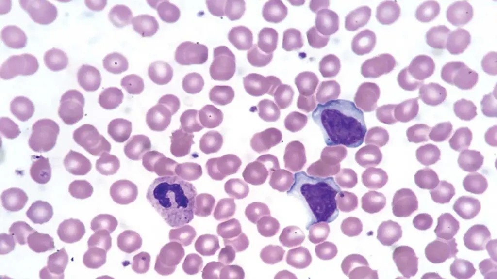
A normal smear shows red cells of similar size and shape with central pallor of about one-third their diameter, platelets scattered between cells, and white cells that, while fewer in number, carry the most diagnostic information.
| Cell Type | Nucleus | Cytoplasm | Granules | Key Features | |
|---|---|---|---|---|---|
| Red Blood Cells | None | Pink to tan, red-orange | None | Biconcave disc; uniform size and central pallor | |
| Neutrophils | Dark purple, 2–5 lobes | Pale pink | Fine lavender to lilac | Segmented mature form | |
| Eosinophils | Blue, bilobed | Blue | Coarse, bright red to red-orange | Distinctive large granules | |
| Basophils | Dark, often obscured | Sky-blue, hidden by granules | Coarse, deep purple to violet-black | Granules dominate the cell | |
| Monocytes | Deep blue-violet, kidney or horseshoe shape | Grey-blue, vacuolated | Fine azurophilic | Largest white cell | |
| Lymphocytes | Round, dense, dark purple | Sky-blue, scant | Usually none | Small cells, thin cytoplasm rim | |
| Platelets | None | None | Violet-purple | Small granular fragments | |
| Malaria Parasites | Red chromatin | Blue cytoplasm | None | Rings and other stages within red cells | |
| Blasts (Leukemia) | Fine chromatin, prominent nucleoli | Variable basophilia |
Variable Auer rods in some myeloblasts |
Immature; high nucleus-to-cytoplasm ratio |
Abnormal findings such as sickle cells, schistocytes, teardrop cells, blasts, and nucleated red cells are graded using ICSH-standardized terminology, which is how modern laboratories report results [3,4].
Troubleshooting
Even experienced technologists get unexpected colors from time to time. The table below maps common problems to causes and fixes.
Wright Stain
| Problem | Common Causes | Solutions |
|---|---|---|
|
Too blue
Excess basophilia
|
Alkaline buffer or rinse water, thick smear, prolonged staining, inadequate rinse | Lower buffer pH toward 6.5, use thinner smears, shorten stain time, rinse adequately |
|
Too pink
Excess eosinophilia
|
Acidic buffer or rinse (often due to unbuffered distilled water absorbing CO₂), prolonged washing, understaining [1] | Adjust buffer pH up toward 6.8, reduce wash time, lengthen stain time |
|
Weak or no staining
Stain failure
|
Exhausted or evaporated stain, too little buffer mixing | Use fresh stain, ensure proper buffer addition and metallic sheen |
|
Precipitate / stain deposit
Artifact
|
Unfiltered stain, stain dried on slide before rinsing | Filter the stain, rinse before the slide dries, keep stain covered |
|
Blue background
Fixation error
|
Inadequate fixation, prolonged storage before fixation, heparin anticoagulant | Fix promptly, use EDTA, refresh smear if old |
|
No metallic sheen on mixing
Ratio issue
|
Incorrect stain-to-buffer ratio | Add more buffer until the sheen forms |
A useful warning
Stain deposits can mimic Gram-positive cocci and lead to false-positive infection reports. Always filter your stain.
Comparison with Other Romanowsky Stains
| Stain | Key Features | Advantages | Disadvantages |
|---|---|---|---|
| Wright | Methanol-based methylene blue + eosin; self-fixing; 3–5 min | Fast, reliable, excellent for white cell differential counts; standard in North America |
Less intense nuclear detail than Giemsa; may not reliably show Plasmodium alone [6] |
| Wright-Giemsa | Wright plus added Giemsa azures | More intense nuclear and basophilic staining; good for bone marrow and parasites |
Slightly longer; more reagents |
| Giemsa | Pure azures + Eosin Y; ~30 min | Best for thick films and bone marrow; vivid color |
Longer protocol; bluish red cells in thin films |
| Leishman | Polychromed methylene blue + eosin in methanol; 15–20 min | Simple; good host-cell contrast in malaria thin films |
Less vivid than Giemsa |
| Field's | Aqueous rapid stain | Very fast (~1 minute); designed for malaria screening |
Less sensitive for thin films |
A comparative study found that Wright stain with deionized water performed equivalently to a modified-Wright control for white cells, platelets, and red cells, supporting its reliability for routine differential analysis [6]. For most teaching and clinical laboratories in North America, the Wright stain is the default first choice, while Wright-Giemsa is preferred when richer nuclear detail or parasite identification is needed.
Safety and Waste Disposal
Wright stain contains methanol, which is flammable and toxic if inhaled, swallowed, or absorbed through the skin. Practical precautions:
- Wear gloves and eye protection. Work in a ventilated space or fume hood.
- Keep stain bottles away from open flames and sparks.
- Collect used stain and rinses in labeled flammable hazardous-waste containers. Do not pour stain waste down the drain or evaporate it.
- Follow your institution's chemical hygiene plan for disposal documentation.
Treat methanol-based stain waste as flammable hazardous waste and let your institutional waste service handle it.
Frequently Asked Questions (FAQs)
What is Wright stain used for?
Wright stain is a Romanowsky-type blood stain used to color a peripheral blood smear so that red cells, white cells, and platelets can be examined under a microscope. It is the standard stain for performing differential white blood cell counts and for evaluating cells in suspected anemia, leukemia, and other blood disorders.
How does Wright stain work?
It uses two oppositely charged dyes. Methylene blue and its azures (basic, blue) bind DNA and RNA, staining nuclei blue-violet. Eosin Y (acidic, orange) binds hemoglobin and basic proteins, staining red cells pink. The methanol solvent fixes the cells, but actual staining begins only after buffer is added and the dyes ionize to form a thiazine-eosinate complex [1].
Why is a buffer added to Wright stain?
Because the staining reaction is pH-dependent. Very little staining happens while undiluted stain sits on the slide. The buffer triggers dye ionization and controls the final color balance. A pH around 6.5–6.8 gives the correct contrast. Too alkaline turns the smear blue; too acidic turns it pink [1,6].
What is the difference between Wright stain and Wright-Giemsa stain?
Wright stain uses methylene blue and eosin. Wright-Giemsa adds Giemsa's azure dyes, producing more intense nuclear and basophilic staining. Wright stain is excellent for routine differential counts, while Wright-Giemsa is often preferred for bone marrow smears and for highlighting parasites.
What does the green "metallic sheen" mean during staining?
It is a visual cue that the stain and buffer have mixed correctly. When you add buffer to the undiluted stain, a green metallic film should appear on the surface. If it does not, add more buffer until it forms.
Is Wright stain safe to use?
The dye itself is generally safe in normal laboratory conditions, but methanol, its solvent, is flammable and toxic if inhaled, swallowed, or absorbed through skin. Always wear gloves and eye protection, work in a ventilated area, and dispose of waste through your institution's flammable hazardous-waste stream rather than down the drain.
Glossary of Related Medical Terms
- Peripheral blood smear (PBS): A thin film of blood spread on a glass slide, stained, and examined under a microscope.
- Romanowsky stain: A family of blood stains that combine a basic dye (methylene blue or its azure derivatives) with an acidic dye (eosin) to color cell components in contrasting shades. Wright, Giemsa, and Leishman stains all belong to this family.
- Polychromatic stain: A stain containing more than one dye component, in this case methylene blue and eosin, producing a range of colors rather than a single shade.
- Methylene blue: A basic, positively charged dye that binds negatively charged structures such as DNA and RNA, staining nuclei blue to violet.
- Eosin Y: An acidic, negatively charged dye that binds positively charged proteins such as hemoglobin, giving red blood cells their pink to tan color.
- Thiazine-eosinate complex: The colored compound formed when methylene blue (or its azure forms) and eosin combine after buffer is added. This complex, not the undiluted stain alone, is what actually colors the cells.
- Polychroming: The chemical process of oxidizing methylene blue into a mixture of azure dyes, broadening the color range of a Romanowsky stain.
- Phosphate buffer: A solution that holds the staining mixture at a stable pH, typically 6.5 to 6.8, so the dyes ionize and bind correctly.
- Metallic sheen: A green, film-like reflection that appears on the slide surface when stain and buffer have mixed in the correct proportion. A practical visual check for proper technique.
- Fixation: A step that preserves cell structure on the slide, usually with methanol, before or during staining.
- Wright-Giemsa stain: A combination stain that adds Giemsa's azure dyes to the standard Wright stain, producing more intense nuclear and basophilic staining. Often preferred for bone marrow smears.
Disclaimer: This protocol is for educational purposes only. Local laboratory standard operating procedures take precedence. It is not intended to be a substitute for informed professional medical advice, diagnosis, or treatment. Always consult a qualified healthcare professional for clinical decision-making. While the information presented here is derived from credible medical sources and is believed to be accurate and up-to-date, it is not guaranteed to be complete or error-free. See additional information.
References
- Keohane, E. M., Preston, M. M., Mirza, K. M., Walenga, J. M. (Eds.). (2024). Rodak's Hematology: Clinical principles and applications (7th ed.). Elsevier.
- Bain, B. J. and Leach, M. (2025). Blood cells: A practical guide (7th ed.). Wiley-Blackwell.
- Palmer, L., Briggs, C., McFadden, S., Zini, G., Burthem, J., Rozenberg, G., Proytcheva, M., & Machin, S. J. (2015). ICSH recommendations for the standardization of nomenclature and grading of peripheral blood cell morphological features. International journal of laboratory hematology, 37(3), 287–303. https://doi.org/10.1111/ijlh.12327
- Zini, G., d'Onofrio, G., Erber, W. N., Lee, S. H., Nagai, Y., Basak, G. W., Lesesve, J. F., & International Council for Standardization in Hematology (ICSH) (2021). 2021 update of the 2012 ICSH Recommendations for identification, diagnostic value, and quantitation of schistocytes: Impact and revisions. International journal of laboratory hematology, 43(6), 1264–1271. https://doi.org/10.1111/ijlh.13682
- ICSH reference method for staining of blood and bone marrow films by azure B and eosin Y (Romanowsky stain). International Committee for Standardization in Haematology. (1984). British journal of haematology, 57(4), 707–710. https://doi.org/10.1111/j.1365-2141.1984.tb02949.x
- Teerasaksilp, S., Wiwanitkit, V., & Lekngam, P. (2005). Comparative study of blood cell staining with wright-giemsa stain, field stain, and a new modified stain. Laboratory hematology : official publication of the International Society for Laboratory Hematology, 11(1), 76–78.