Key Takeaways
Leukopenia is a total white blood cell count below 4,000 cells per microliter (< 4 x 10⁹/L) in adults. It signals a weakened defense against infection, not a disease in itself.
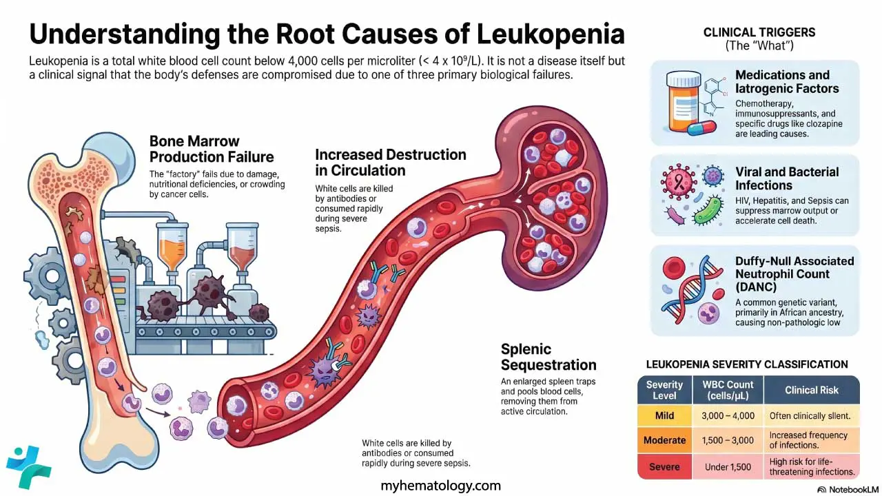
- Classification ▾: Mild leukopenia (3,000–4,000), moderate (1,500–3,000), and severe (<1,500) carry progressively higher infection risk. An ANC under 500 (< 0.5 x 10⁹/L) places a patient at high risk for serious bacterial and fungal infection.
- Causes ▾: Reduced bone marrow production, increased destruction in the circulation, and sequestration in organs such as the spleen. Chemotherapy, viral infection, and autoimmune disease are the most common triggers.
- Symptoms ▾: Often subtle, primarily related to increased infection risk (frequent/severe infections, fever, sore throat, etc.) and sometimes underlying cause.
- Laboratory Investigations ▾: CBC with differential is key; peripheral blood smear, bone marrow study, infection screens, and autoimmune panels follow as needed.
- Treatment & Management ▾: Treatment targets the underlying cause while supportive care prevents infection. Fever in a neutropenic patient is a medical emergency [1,4].
*Click ▾ for more information
Introduction
Leukopenia means having fewer white blood cells than normal. Because white blood cells are the immune system's front-line defenders, leukopenia can leave the body open to infection. The condition itself is usually silent. Patients often discover it on a routine blood test or because they keep catching infections that won't quit.
This article walks through what leukopenia is, why it happens, how clinicians investigate it, and how it is managed.
What is Leukopenia (Low White Cell Count)?
In adults, the white blood cell count usually sits between 4,000 and 11,000 cells per microliter of blood (or 4.0–11.0 × 10⁹/L in SI units). Children's ranges differ and trend higher in infancy. Counts naturally vary with age, ethnicity, smoking status, time of day, and medications.
Leukopenia is defined as a total WBC under 4,000/µL.
Classification of severity
- Mild leukopenia: 3,000–4,000/µL (3.0–4.0 × 10⁹/L). Often clinically silent.
- Moderate leukopenia: 1,500–3,000/µL (1.5–3.0 × 10⁹/L). Infections become more frequent or harder to clear.
- Severe leukopenia: Under 1,500/µL (1.5 × 10⁹/L). Serious and sometimes life-threatening infections are a real risk.
Why neutrophils matter most
Neutrophils make up roughly 60% of circulating white cells. They are the cells that swarm bacterial and fungal invaders. So when total WBC drops, what really drives infection risk is how far the neutrophil count drops with it. That number is the absolute neutrophil count (ANC).

Neutropenia severity by ANC
How to calculate the ANC
ANC = Total WBC × (% segmented neutrophils + % bands) ÷ 100
Worked example: A patient has a WBC of 2,000/µL with 30% neutrophils. ANC = 2,000 × 30 ÷ 100 = 600/µL → moderate neutropenia.
Absolute Neutrophil Count (ANC)
The role of lymphocytes. While neutrophils fight bacteria, lymphocytes are crucial for viral and opportunistic defense. A low Absolute Lymphocyte Count (ALC) which are common in severe viral infections, advanced HIV, or after treatment with biologic drugs like rituximab can also drive leukopenia and requires its own independent monitoring [8].
A note on Duffy-null associated neutrophil count
Not every low count is pathologic. A common genetic variant in the ACKR1 gene (the Duffy-null genotype) produces a lifelong, mildly low neutrophil count without raising infection risk. The variant is highly prevalent in people of African ancestry (around 80%) and Middle Eastern ancestry (around 25%), and far less common in European and East Asian populations [6].
Hematology bodies increasingly use the term Duffy-null associated neutrophil count (DANC) in place of the older "benign ethnic neutropenia" [6]. Recognizing this variant matters: misclassifying it as a disease can block patients from chemotherapy, clozapine, and other treatments that have neutrophil thresholds.
Causes of Leukopenia
Three mechanisms underpin almost every case: the marrow isn't making enough white cells, the cells are being destroyed once they reach the blood, or they are being trapped somewhere they shouldn't be.
1. Bone marrow can't produce enough cells
The bone marrow is the factory floor for blood cells. Damage there shows up in the blood.
- Aplastic anemia: The marrow shuts down across all three blood lines. Causes include autoimmune attack, certain infections, toxins, radiation, or no identifiable trigger.
- Myelodysplastic syndromes (MDS): The marrow produces blood cells that don't mature properly.
- Leukemia, lymphoma, multiple myeloma: Cancer cells crowd out normal hematopoiesis (blood-cell production).
- Myelofibrosis: Scar tissue replaces marrow.
- Congenital syndromes: Fanconi anemia, Diamond-Blackfan anemia, and severe congenital neutropenia present in childhood.
- Nutritional deficiencies: Vitamin B12, folate, and copper are essential for DNA synthesis in dividing marrow cells. Without them, blood-cell production stalls.
2. White cells are destroyed in circulation
- Autoimmune neutropenia: The immune system makes antibodies that target neutrophils.
- Drug-induced immune destruction: Specific drugs trigger antibody-mediated killing of white cells.
- Severe sepsis: Cells are consumed faster than the marrow can replace them.
3. White cells are sequestered (trapped)
- Hypersplenism: An enlarged spleen pools and destroys blood cells. Common in liver cirrhosis and portal hypertension.
Infections
Some infections both fight and lower white cells.
- Viruses: HIV, hepatitis viruses, Epstein-Barr virus, cytomegalovirus, influenza, and parvovirus B19 commonly suppress marrow output or accelerate destruction.
- Bacteria: Typhoid, brucellosis, and overwhelming sepsis can do the same.
- Other: Malaria and miliary tuberculosis.
Medications
The list is long. The most clinically important:
- Chemotherapy: Kills rapidly dividing marrow cells along with the cancer. Counts typically reach their lowest point (the nadir) 7–14 days after a cycle.
- Cellular and Immunotherapies: Chimeric antigen receptor (CAR) T-cell therapies and bispecific antibodies often cause profound, atypical cytopenias that can persist for weeks to months, requiring extended monitoring well beyond the traditional chemotherapy nadir [9].
- Immunosuppressants: Azathioprine, methotrexate, mycophenolate, cyclosporine.
- Antibiotics: Trimethoprim-sulfamethoxazole most notably.
- Antiseizure drugs: Carbamazepine, valproate, and others can lower counts; pediatric studies show this is usually mild and rarely changes management [3].
- Psychotropics: Clozapine carries a specific, well-known risk of severe neutropenia and requires monitored prescribing.
- NSAIDs: Rare but recognized.
Autoimmune disorders
- Systemic lupus erythematosus (SLE): Leukopenia is part of the classification criteria.
- Felty's syndrome: Rheumatoid arthritis with splenomegaly and neutropenia.
Other contributors
Radiation therapy directed at marrow-rich bones, major surgery or trauma, and hemodialysis can all transiently lower counts.
Symptoms of Leukopenia
Leukopenia itself doesn't usually cause symptoms. What people notice are the infections a weakened immune system fails to stop, plus signs of whatever disease is causing the low count in the first place.
Signs that infection is taking hold
- Fever or chills
- Sore throat, mouth ulcers
- Cough, breathlessness
- Burning urination
- Skin redness, warmth, or pus at a wound site
- Diarrhea
- Persistent fatigue
A pattern of frequent, recurrent, prolonged, or unusually severe infections is the classic clinical clue. In severe leukopenia, opportunistic infections (caused by microbes that wouldn't trouble a healthy immune system) become possible.
Signs of the underlying cause
These vary with the driver of the leukopenia.
- Bone pain, easy bruising, or bleeding → think marrow disorder.
- Swollen lymph nodes, night sweats, weight loss → think lymphoma or chronic infection.
- Skin rash, joint pain → think autoimmune disease.
- Pale skin → think coexisting anemia.
- Enlarged spleen or liver → think hypersplenism, leukemia, or lymphoma.
Laboratory Investigations
The workup answers two questions: how low is the count, and why?
Step 1: Confirm and characterize
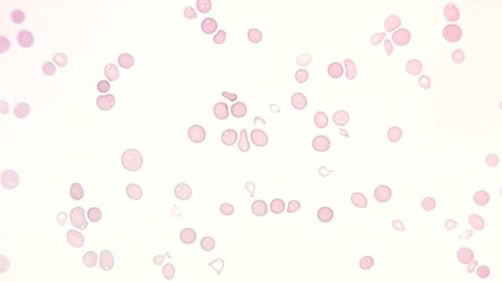
Complete blood count with differential. This is the foundation. It gives the total WBC, the breakdown by cell type, the hemoglobin, and the platelet count. The differential reveals whether neutropenia, lymphopenia, or both are driving the picture.
Peripheral blood smear. A trained eye on the slide can spot blast cells (suggesting leukemia), atypical lymphocytes (viral infection), dysplastic features (MDS), or coexisting red-cell and platelet abnormalities. The smear catches qualitative changes the automated counter misses.
Step 2: Find the cause
Bone marrow aspiration and biopsy. Often the definitive test. Aspiration samples the liquid marrow for cellular detail; biopsy provides a tissue core for architecture. Add-on studies like flow cytometry, cytogenetics, molecular panels can clarify malignancies and inherited syndromes.
Infection screen. Blood cultures, viral serologies (HIV, EBV, CMV, hepatitis, parvovirus B19), and PCR assays as the clinical picture demands.
Autoimmune workup. ANA, anti-dsDNA, anti-Sm, rheumatoid factor, ANCA, and a direct antiglobulin test when indicated.
Nutritional studies. Vitamin B12, folate, copper, and iron studies.
Drug review. A careful list of prescription drugs, over-the-counter agents, and supplements. Stopping a suspect drug and watching the count rebound is often the diagnostic test.
Imaging. Ultrasound or CT for splenomegaly; chest imaging for pulmonary infection; lymph-node biopsy if lymphadenopathy points to lymphoma.
Genetic testing. When a congenital syndrome is suspected, particularly in pediatric patients.
Treatment and Management of Leukopenia
Treatment has two parallel tracks: fix the underlying cause, and protect the patient from infection while counts recover.
Treating the underlying cause
- Infection: Targeted antimicrobial therapy. Counts typically rebound once the pathogen is cleared.
- Medication-induced leukopenia: Stop the offending drug if clinically possible. Monitor recovery. Substitute when needed.
- Autoimmune disease: Treat the parent condition with corticosteroids, immunosuppressants, or biologic agents.
- Nutritional deficiency: Replace the missing nutrient — oral or intramuscular B12, oral folate, copper as indicated.
- Hematologic malignancies and marrow failure: Treatment depends on the diagnosis and may include chemotherapy, targeted therapy, immunosuppression (often combined with eltrombopag in aplastic anemia), or allogeneic stem cell transplant.
- Hypersplenism: Address the underlying liver or hematologic cause. Splenectomy is reserved for selected cases.
Supportive care: preventing infection
- Hand hygiene. The single most effective measure for the patient, family, and clinical staff.
- Avoid sick contacts and crowded spaces during periods of significant neutropenia.
- Safe food practices. Thoroughly cooked meat, washed produce, no unpasteurized dairy or raw seafood.
- Mouth and skin care. Gentle brushing with a soft toothbrush, prompt cleaning of any break in the skin.
- Vaccination, with care. Inactivated vaccines (annual influenza, pneumococcal, COVID-19) are encouraged when timed appropriately. Live vaccines like MMR, varicella, yellow fever, BCG, live-attenuated influenza are generally contraindicated during significant immunosuppression.
Managing fever in a neutropenic patient
This is the highest-stakes scenario in leukopenia care. Operational definitions used worldwide are clear: neutropenia for this purpose is an ANC under 500/µL, or under 1,000/µL with an expected fall below 500 within 48 hours. Neutropenic fever is a single oral temperature of 38.3°C (101°F), or 38.0°C (100.4°F) sustained for more than one hour, in such a patient [4].
A neutropenic fever triggers a same-day, often within-the-hour, response:
- Blood cultures from peripheral blood and any indwelling lines.
- Urine and other site-specific cultures.
- Empiric broad-spectrum antibiotic monotherapy with an anti-pseudomonal beta-lactam (commonly piperacillin-tazobactam, cefepime, or meropenem) [4].
- Risk stratification for outpatient care using validated tools like the MASCC or CISNE scores identify low-risk patients. Rather than automatic admission for IV therapy, appropriately selected low-risk patients can often be safely managed in the outpatient setting with oral antibiotics (typically ciprofloxacin plus amoxicillin-clavulanate) [10].
- If fever persists beyond 4–7 days in a high-risk patient, empiric antifungal therapy is added [4].
Growth factors
Granulocyte colony-stimulating factors (G-CSFs) push the marrow to produce more neutrophils. They are most often used after specific chemotherapy regimens, in severe or febrile neutropenia, and in selected congenital syndromes.
- Filgrastim (short-acting, daily injection)
- Pegfilgrastim (long-acting, once per chemotherapy cycle — now standard in many regimens)
- Sargramostim (GM-CSF, narrower use)
Granulocyte transfusion
Rarely used. Reserved for severe, refractory bacterial or fungal infection in profoundly neutropenic patients when nothing else is working. The brief lifespan of transfused white cells and the risk of transfusion reactions limit its role.
Monitoring
Serial CBCs track recovery. The frequency depends on severity, the cause, and the treatment phase. Patients on chemotherapy typically have counts checked weekly, with closer monitoring around the expected nadir.
Potential Complications
The lower the count and the longer it lasts, the higher the stakes.
- Bacterial infections are the most immediate threat. Skin, urinary, respiratory, and bloodstream infections can escalate fast in a neutropenic patient.
- Fungal infections (Candida, Aspergillus) become an issue with profound, prolonged neutropenia [4].
- Viral reactivation of herpes simplex, varicella zoster, or CMV can occur.
- Opportunistic infections such as Pneumocystis jirovecii pneumonia emerge with severe immune suppression.
- Sepsis and septic shock. A neutropenic patient with fever can deteriorate within hours. Fever in this context is treated as sepsis until proven otherwise.
- Delayed wound healing because neutrophils and macrophages are central to tissue repair.
- Mucositis — painful inflammation of the mouth and gut lining — is common with chemotherapy and worsens infection risk.
- Progression of the underlying disease. MDS can transform to acute myeloid leukemia; untreated HIV progresses to AIDS-defining infections.
Living with Leukopenia: Notes for Patients and Caregivers
Practical day-to-day steps make a real difference. Wash hands often. Carry a thermometer and check the temperature at the first hint of feeling unwell. Know the phone number for the on-call hematology or oncology team and use it for any fever — do not "wait and see." Keep a current medication list. Eat well-cooked food and skip the salad bar during nadir periods. Ask the team in advance which vaccines are safe and when they should be given relative to chemotherapy cycles. Most patients can live full lives between cycles or between flares. The key is recognizing red flags early.
Conclusion
Leukopenia is a finding on a blood count, not a single disease. Understanding the underlying mechanism (production failure, destruction, or sequestration) guides every subsequent step from investigation to treatment. The absolute neutrophil count, not the total WBC, drives most clinical decisions. And while severe leukopenia carries real risk, modern supportive care, growth factors, and prompt management of febrile neutropenia have transformed outcomes [4,5].
Key Takeaway: Recognize the signs, act early on fever, and treat the cause.
Frequently Asked Questions (FAQs)
What WBC count is considered leukopenia?
In adults, leukopenia means a total white blood cell count below 4,000 cells per microliter of blood. Doctors also check the absolute neutrophil count (ANC), because neutrophils are the cells most responsible for fighting bacterial and fungal infection. An ANC under 1,500 is neutropenia; under 500 is severe and carries the highest infection risk.
Is leukopenia the same as neutropenia?
No, but they overlap. Leukopenia is a drop in total white blood cells. Neutropenia is a drop in one specific type, the neutrophil. Because neutrophils make up most white cells, most cases of leukopenia are driven by neutropenia. Clinicians focus on the ANC because that number predicts infection risk better than the total WBC.
What is the most common cause of leukopenia?
It depends on the setting. In cancer wards, it's chemotherapy. In primary care, viral infections (influenza, EBV, hepatitis, HIV) are the usual suspects. Medications, autoimmune disease, and nutritional deficiencies are other common causes. A low count in someone of African, Middle Eastern, or West Indian ancestry may also reflect the benign Duffy-null genotype, not disease [6].
When does leukopenia become a medical emergency?
Fever in a neutropenic patient is a medical emergency. The threshold is a single oral temperature of 38.3°C (101°F), or 38.0°C (100.4°F) sustained for more than an hour, in someone with an ANC under 500 (or expected to fall below 500). This is febrile neutropenia. It needs immediate evaluation, blood cultures, and broad-spectrum antibiotics within an hour [4].
Can leukopenia be cured?
It depends on the cause. Viral or drug-related leukopenia usually resolves on its own. Chemotherapy-induced leukopenia recovers as the marrow rebounds over a few weeks. Genetic, autoimmune, and marrow-failure causes are managed long-term rather than cured. The Duffy-null variant requires no treatment.
How can someone with leukopenia lower their infection risk at home?
Wash hands often. Avoid crowds and people with active infections. Cook meat thoroughly, wash produce, and skip raw or unpasteurized foods. Maintain meticulous mouth and skin care. Ask the healthcare team which vaccines are safe — live vaccines such as MMR, varicella, and yellow fever are generally off-limits during significant immunosuppression.
Glossary of Related Medical Terms
- Leukocyte: A white blood cell. Part of the immune system.
- Leukopenia: A total white blood cell count below the normal range, usually under 4,000 cells per microliter in adults.
- Neutrophil: The most common white blood cell. Fights bacteria and fungi.
- Neutropenia: A low neutrophil count. Usually defined as an absolute neutrophil count under 1,500 cells per microliter.
- Absolute Neutrophil Count (ANC): The actual number of neutrophils per microliter of blood. Calculated from the total WBC and the differential.
- Differential count: The breakdown of the total WBC into its subtypes (neutrophils, lymphocytes, monocytes, eosinophils, basophils).
- Bone marrow: The spongy tissue inside bones where blood cells are made.
- Hematopoiesis: The process of making new blood cells.
- Myelosuppression: A drop in bone marrow activity, often caused by chemotherapy or radiation. Results in fewer blood cells of all types.
- Pancytopenia: A low count of all three blood cell lines: red cells, white cells, and platelets.
- Phagocytosis: The process by which a cell engulfs and digests a microbe.
- Demargination: The shift of neutrophils from the vessel wall back into the flowing blood, raising the measured count.
- Sequestration: Trapping of blood cells in an organ such as the spleen.
- Opportunistic infection: An infection caused by a microbe that does not usually cause disease in people with a normal immune system.
- Mucositis: Painful inflammation and ulceration of the lining of the mouth or gut, often from chemotherapy.
- Febrile neutropenia: Fever in a patient with neutropenia. A medical emergency.
- Nadir: The lowest point a blood count reaches after chemotherapy, usually 7–14 days post-treatment.
- G-CSF: Granulocyte colony-stimulating factor. A medication (filgrastim, pegfilgrastim) that prompts the bone marrow to make more neutrophils.
- Duffy-null genotype: A genetic variant common in people of African, Middle Eastern, and West Indian ancestry. Linked to a naturally lower neutrophil count without raised infection risk.
Disclaimer: This article is intended for informational purposes only and is specifically targeted towards medical students. It is not intended to be a substitute for informed professional medical advice, diagnosis, or treatment. While the information presented here is derived from credible medical sources and is believed to be accurate and up-to-date, it is not guaranteed to be complete or error-free. See additional information.
References
- Shoenfeld, Y., Alkan, M. L., Asaly, A., Carmeli, Y., & Katz, M. (1988). Benign familial leukopenia and neutropenia in different ethnic groups. European journal of haematology, 41(3), 273–277. https://doi.org/10.1111/j.1600-0609.1988.tb01192.x
- Ing, V. W. (1984). The etiology and management of leukopenia. Canadian Family Physician, 30, 1835–1839.
- Pettersson, H., Alani, T., Rydén, I., Stödberg, T., Eksborg, S., & Sundin, M. (2025). Leukopenia in Children on Antiseizure Medication is Common and Has Minor Clinical Impact. The Journal of pediatrics, 282, 114593. https://doi.org/10.1016/j.jpeds.2025.114593
- Giamarellou, H., & Antoniadou, A. (2001). Infectious complications of febrile leukopenia. Infectious disease clinics of North America, 15(2), 457–482. https://doi.org/10.1016/s0891-5520(05)70156-2
- Adamietz, I. A., Rosskopf, B., Dapper, F. D., von Lieven, H., & Boettcher, H. D. (1996). Comparison of two strategies for the treatment of radiogenic leukopenia using granulocyte colony stimulating factor. International journal of radiation oncology, biology, physics, 35(1), 61–67. https://doi.org/10.1016/s0360-3016(96)85012-7
- Merz, L. E., Story, C. M., Osei, M. A., Jolley, K., Ren, S., Park, H. S., Yefidoff Freedman, R., Neuberg, D., Smeland-Wagman, R., Kaufman, R. M., & Achebe, M. O. (2023). Absolute neutrophil count by Duffy status among healthy Black and African American adults. Blood advances, 7(3), 317–320. https://doi.org/10.1182/bloodadvances.2022007679
- Strasfeld, L. (2025). Febrile neutropenia. BMJ Best Practice.
- Merck Manual Professional Version. (2023). Lymphocytopenia. Merck & Co., Inc. Retrieved from https://www.msdmanuals.com/professional/hematology-and-oncology/leukopenias/lymphocytopenia[
- Jain, T., Olson, T. S., & Locke, F. L. (2023). How I treat cytopenias after CAR T-cell therapy. Blood, 141(20), 2460–2469. https://doi.org/10.1182/blood.2022017415
- Taplitz, R. A., Kennedy, E. B., & Flowers, C. R. (2018). Outpatient Management of Fever and Neutropenia in Adults Treated for Malignancy: American Society of Clinical Oncology and Infectious Diseases Society of America Clinical Practice Guideline Update Summary. Journal of oncology practice, 14(4), 250–255. https://doi.org/10.1200/JOP.18.00016


