Key Takeaways
Anemia is a sign, not a diagnosis. A systematic approach to anemia means finding the underlying cause before treating it, because iron, B12, kidney disease, inflammation, and inherited disorders each need a different plan.
- Detailed Clinical History ▾: Comprehensive clinical history requires evaluating the patient's presenting symptoms, past medical and medication records, lifestyle factors including diet and social habits, menstrual history, and family background to accurately identify underlying conditions such as anemia.
- Physical Examination ▾: A comprehensive physical examination involves evaluating vital signs, checking for visible indicators like pallor or jaundice, conducting a full systemic assessment, and performing a targeted neurological exam if a specific deficiency is suspected.
- Laboratory Investigations ▾: The CBC, MCV, and reticulocyte count together drive the workup. Microcytic anemias point to iron deficiency or thalassemia; macrocytic anemias point to B12 or folate deficiency; reticulocytes tell you whether the marrow is responding.
- Differential Diagnosis ▾: Based on clinical findings and laboratory tests, healthcare professionals create a list of possible causes of anemia based on the MCV value (microcytic, normocytic, macrocytic).
- Treatment ▾: The approach emphasizes treating the underlying cause of anemia. This can involve iron supplementation, vitamin B12 injections, blood transfusions, or managing chronic diseases that contribute to anemia.
*Click ▾ for more information
Introduction
Anemia is one of the most common conditions you will meet in clinical practice. It affects roughly 1.92 billion people worldwide, or about a quarter of the global population, and the burden falls hardest on women and young children [2]. Yet anemia is not a diagnosis on its own. It is a signal that something else is going wrong, from a slow gastrointestinal bleed to an inherited hemoglobin disorder.
A systematic approach to anemia is how clinicians turn that signal into a specific cause and a targeted plan. The goal is simple: find out why hemoglobin is low, fix the underlying problem, and avoid unnecessary tests along the way.
This article walks through that process by providing a clear definition first, then the history, examination, and lab tests that build the diagnosis, and finally the modern treatment options.
What anemia actually means
Anemia is a reduction in hemoglobin (the oxygen-carrying protein inside red blood cells) or in red cell mass below the normal range for a person's age, sex, and physiological state. Less hemoglobin means less oxygen reaches tissues. That is why patients feel tired, look pale, and breathe faster than expected.
The World Health Organization sets the diagnostic cut-offs. In March 2024, WHO updated several of them, including the threshold for children aged 6–23 months (now 10.5 g/dL), the second-trimester pregnancy threshold (now 10.5 g/dL), and the way hemoglobin is adjusted for altitude (starting at 500 m above sea level instead of 1,000 m) and smoking [1].
Why systematic matters
Skipping steps is the most common reason anemia is misdiagnosed. A patient labeled as "iron deficient" without iron studies may actually have anemia of inflammation, where iron supplements will not help and may delay the real diagnosis. A clean, repeatable workflow protects both the patient and your reasoning. The five steps below — history, examination, focused labs, differential, and targeted treatment — are that workflow.
Step 1: Take a Detailed Clinical History
History is where most diagnoses are made. Before any blood is drawn, a careful conversation usually narrows the cause to a handful of possibilities.
History of the presenting illness
Ask about onset, duration, and severity. Anemia that builds over months feels different from anemia that appears in a week. Cover the classic symptoms:
- Fatigue, weakness, breathlessness on exertion
- Dizziness, headache, palpitations
- Pale skin, cold extremities, poor concentration
- Any recent or ongoing blood loss: heavy periods, black or bloody stools, surgery, or trauma
Menstrual history
In any menstruating patient, ask about cycle regularity, duration, and how heavy the flow is. Heavy menstrual bleeding is one of the leading causes of iron deficiency in women of reproductive age.
Past medical, surgical, and drug history
Chronic kidney disease, autoimmune disorders, inflammatory bowel disease, and previous gastrointestinal surgery (especially gastrectomy or ileal resection) all matter. Ask about medications too: NSAIDs and aspirin can cause occult GI bleeding; metformin and proton pump inhibitors interfere with vitamin B12 absorption; some antibiotics and chemotherapy agents suppress the bone marrow.
Social and dietary history
A short conversation about diet, alcohol, and travel often surfaces the cause. Vegans and strict vegetarians without B12 supplementation are at risk of deficiency. Heavy alcohol use damages the marrow and impairs folate absorption. Travel to malaria-endemic regions or hookworm-prevalent areas changes the differential.
Ask about pica — cravings for ice, clay, or dirt. It is a surprisingly specific clue to iron deficiency.
Family history
Sickle cell disease, thalassemia, hereditary spherocytosis, and G6PD deficiency all run in families. A grandmother with "anemia all her life" is a hint worth chasing.
Epidemiology of Anemia
Understanding the epidemiology of anemia helps identify individuals at higher risk and guide appropriate screening strategies.
Age and Sex Predilection
- Iron Deficiency Anemia (IDA): Most common globally, affecting:
- Children: Particularly prevalent in preschoolers due to rapid growth, inadequate iron intake, and potential blood loss from parasitic infections.
- Pregnant Women: Increased iron demands during pregnancy put them at risk, especially with inadequate dietary intake or multiple pregnancies.
- Adolescent Girls and Women of Reproductive Age: Heavy menstrual bleeding is a significant risk factor.
- Vitamin B12 and Folate Deficiency Anemia: More common in older adults due to:
- Reduced dietary intake and absorption issues.
- Use of medications that interfere with B12 absorption (metformin, proton pump inhibitors).
- Anemia of Chronic Disease (ACD): Affects individuals with underlying chronic conditions like:
- Kidney disease (all age groups).
- Autoimmune diseases (more common in women).
- Cancer (all age groups).
Geographical Variations
- Iron Deficiency Anemia: More prevalent in developing countries due to:
- Poverty and limited access to iron-rich foods.
- High prevalence of parasitic infections that contribute to blood loss.
- Vitamin and Mineral Deficiencies: More common in areas with limited access to fortified foods and diverse diets.
- Infectious Diseases: Malaria, hookworm, and other chronic infections can contribute to anemia in certain regions.
- Genetic Hemoglobinopathies: Sickle cell disease and thalassemia have specific geographical distributions. Sickle cell disease has a higher prevalence in individuals of African descent. Alpha thalassemia is common in Southeast Asia and the Mediterranean. Beta thalassemia is more prevalent in the Middle East, Southeast Asia, and the Mediterranean.
Risk Factors
- Dietary Habits: Diets low in iron, vitamin B12, and folate increase the risk of deficiency anemia. Vegetarian or vegan diets without proper planning can be a risk factor.
- Medications: Certain medications can suppress bone marrow function or interfere with nutrient absorption, leading to anemia.
- Chronic Medical Conditions: Kidney disease, autoimmune disorders, and inflammatory bowel disease can contribute to anemia.
- Heavy Menstrual Bleeding: Can lead to iron deficiency anemia in women.
- Pregnancy: Increased iron demands during pregnancy put women at risk.
- Gastrointestinal Issues: Conditions like ulcers or celiac disease can impair nutrient absorption.
- Alcohol Abuse: Damages bone marrow and interferes with vitamin B12 absorption.
- Recent Surgery: Blood loss during major surgeries can contribute to anemia.
- Certain Cancers: Can suppress bone marrow function and lead to anemia.
By understanding these epidemiological factors, healthcare professionals can identify populations at higher risk for specific types of anemia.
Step 2: Perform a Focused Physical Examination
The physical exam confirms suspicions and adds new ones.
General inspection
Look at the conjunctiva, palms, and tongue for pallor. Check the sclerae for jaundice (suggests hemolysis or liver disease). Look at the nails for koilonychia (spoon-shaped, in chronic iron deficiency) and the tongue for glossitis (smooth, sore tongue, in B12 or iron deficiency).
Vital signs
Tachycardia and tachypnea are the body's compensation for low oxygen-carrying capacity. The heart pumps faster and the lungs breathe faster to deliver the same amount of oxygen with fewer red cells. Hypotension may appear in severe or acute blood loss.
System-by-system
- Cardiovascular: A flow murmur (soft, systolic) is common in significant anemia.
- Abdomen: Feel for splenomegaly — an enlarged spleen suggests hemolysis, thalassemia, or hematological malignancy.
- Lymph nodes: Enlargement raises concern for infection or malignancy.
- Neurological: In suspected B12 deficiency, check vibration sense and proprioception. Subacute combined degeneration of the spinal cord can occur even before macrocytosis appears.
Step 3: Order the Right Laboratory Investigations
Tests should follow the history, not replace it. The complete blood count (CBC) is the foundation; everything else is targeted.
Complete Blood Count (CBC)
The CBC tells you not just whether the patient is anemic, but what kind of anemia they have. Three numbers do most of the work:
- Hemoglobin (Hb): Confirms anemia using the 2024 WHO thresholds [1].
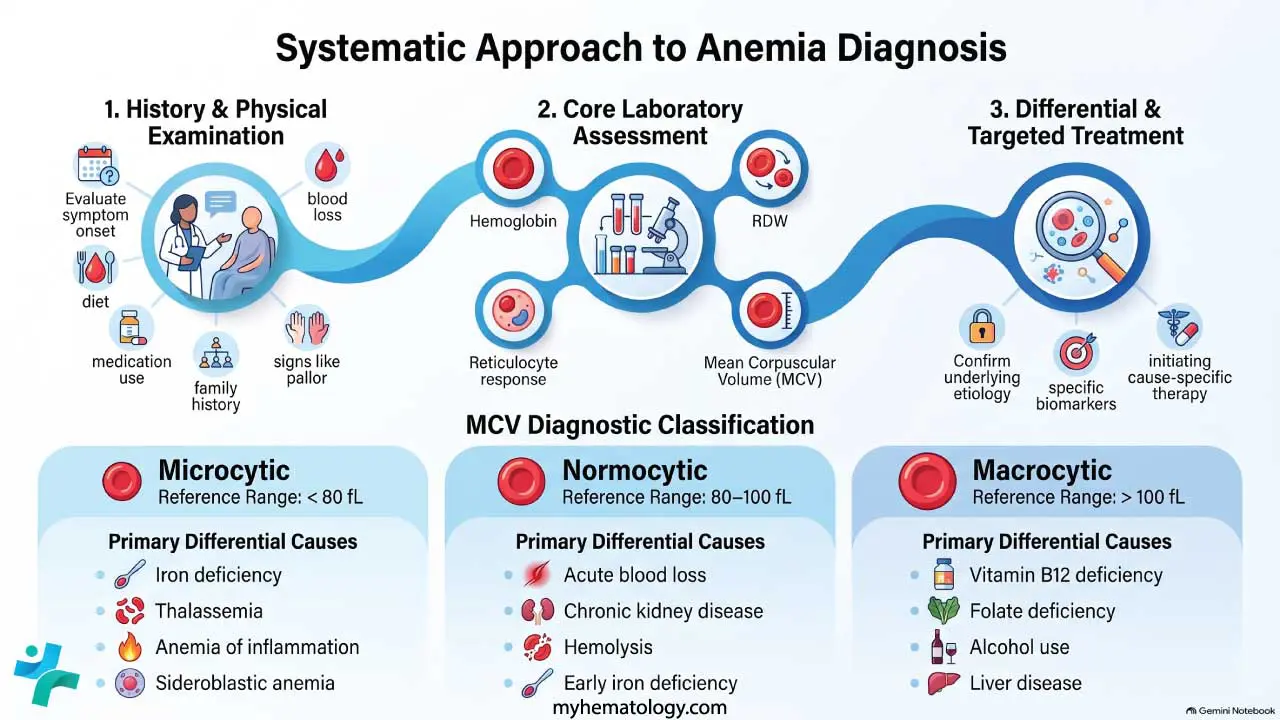
- Mean corpuscular volume (MCV): Sorts anemia into microcytic, normocytic, or macrocytic.
- Red cell distribution width (RDW): A high RDW points to mixed causes, such as combined iron and B12 deficiency.
MCV and the classic categories
| MCV | Category | Common Causes |
|---|---|---|
| < 80 fL | Microcytic | Iron deficiency, thalassemia, anemia of inflammation, sideroblastic anemia |
| 80–100 fL | Normocytic | Acute blood loss, early iron deficiency, anemia of chronic kidney disease, hemolysis, aplastic anemia, mixed deficiencies |
| > 100 fL | Macrocytic | Vitamin B12 deficiency, folate deficiency, alcohol use, liver disease, hypothyroidism, myelodysplastic syndromes |
The reticulocyte count: don't skip it
The reticulocyte count tells you how the bone marrow is responding. A high count means the marrow is making extra red cells, usually because they are being lost (bleeding) or destroyed (hemolysis). A low or inappropriately normal count means the marrow is failing to keep up for example, through deficiency, inflammation, kidney disease, or marrow disease itself. Pairing MCV with the reticulocyte response is how anemia is now taught and how clinicians actually think.
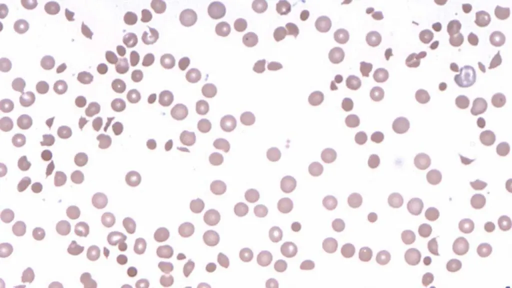
The peripheral blood smear
A smear is a free, fast, and surprisingly informative test. It can reveal:
- Microcytic, hypochromic cells (iron deficiency, thalassemia)
- Macro-ovalocytes and hypersegmented neutrophils (B12 or folate deficiency)
- Schistocytes (microangiopathic hemolytic anemia)
- Sickle cells (sickle cell disease)
- Target cells (thalassemia, liver disease)
- Spherocytes (hereditary spherocytosis or autoimmune hemolysis)
Iron Studies
The four-test panel of serum iron, ferritin, transferrin saturation, and TIBC distinguishes iron deficiency from anemia of inflammation. Two patterns to memorize:
- Iron deficiency: low ferritin, low transferrin saturation, high TIBC. Ferritin < 30 µg/L is the most specific finding [6].
- Anemia of inflammation: normal-to-high ferritin (because ferritin rises with inflammation), low transferrin saturation, low or normal TIBC. The mechanism is hepcidin, a hormone that locks iron inside storage cells when the body senses inflammation [7].
This is why a patient with rheumatoid arthritis can have plenty of iron but still be anemic as the iron just cannot leave the storage compartment to reach the marrow.
B12 and folate
Order both together. Low B12 needs follow-up testing for the cause, especially pernicious anemia (an autoimmune destruction of intrinsic factor-producing cells in the stomach), which is the most common reason for B12 deficiency in older adults.
Targeted further tests
Depending on the picture, you may add:
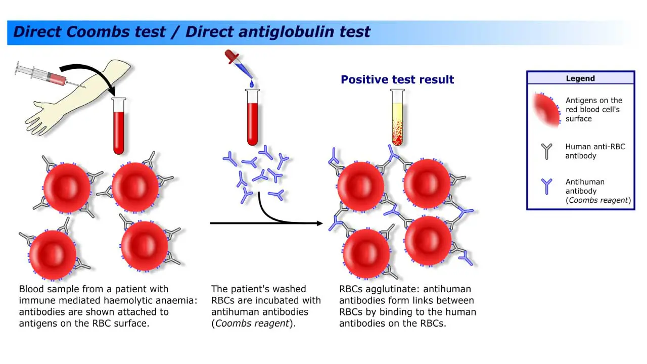
- Hemolysis panel: elevated LDH, low haptoglobin, elevated indirect bilirubin, plus the direct antiglobulin test (DAT/Coombs) to look for immune-mediated destruction
- Hemoglobin electrophoresis for thalassemia and sickle cell disease
- Stool occult blood and endoscopy for suspected GI bleeding
- Kidney and liver function tests
- Bone marrow examination in unexplained pancytopenia or suspected primary marrow disease
Step 4: Build the Differential Diagnosis
Combine the MCV, the reticulocyte response, and the clinical picture. A young woman with heavy periods, microcytic anemia, low ferritin, and high TIBC has iron deficiency until proven otherwise. An older patient with macrocytic anemia, neurological symptoms, and a strict vegan diet probably has B12 deficiency. A child of Mediterranean ancestry with persistent microcytosis and normal iron studies needs hemoglobin electrophoresis for thalassemia.
The diagnosis should always fit the patient and not just the lab numbers.
Step 5: Treat the Underlying Cause
Modern anemia care is more than iron tablets and transfusions. Treatment now depends heavily on cause and severity.
Iron deficiency anemia
- Oral iron (ferrous sulfate, fumarate, or gluconate) remains first-line. Alternate-day dosing improves absorption and reduces side effects [6].
- Intravenous iron (ferric carboxymaltose, ferric derisomaltose, ferumoxytol) is now safer and faster than older preparations. It is used when oral iron fails, is not tolerated, or when rapid replenishment is needed (pregnancy, pre-surgery, inflammatory bowel disease).
- Always treat the cause of the loss — investigate menstrual or GI bleeding rather than just refilling the tank [6].
Vitamin B12 and folate deficiency
B12 is replaced with intramuscular injections or high-dose oral therapy; pernicious anemia generally requires lifelong replacement. Folate deficiency is corrected with oral folic acid. Always confirm B12 status before giving folate alone as folate can mask B12 deficiency while neurological damage progresses.
Anemia of chronic kidney disease
Erythropoiesis-stimulating agents (epoetin alfa, darbepoetin) have been the standard for decades. Newer HIF-prolyl hydroxylase inhibitors such as roxadustat and daprodustat are now approved in many countries as oral alternatives, with comparable efficacy in non-dialysis CKD patients [3]. Iron stores must be repleted alongside.
Anemia of inflammation
Treat the underlying inflammatory or chronic disease. Iron supplementation alone often does not work because hepcidin is blocking iron release [7].
Sickle cell disease
The treatment landscape has changed substantially. Hydroxyurea remains foundational, but the modern toolkit also includes L-glutamine, voxelotor (which increases hemoglobin's oxygen affinity), and crizanlizumab (which reduces vaso-occlusive crises). In December 2023, the FDA approved two gene therapies — exagamglogene autotemcel (Casgevy, the first CRISPR-based therapy) and lovotibeglogene autotemcel (Lyfgenia) — for severe sickle cell disease [4].
Thalassemia
Severe forms still rely on regular transfusions and iron chelation to prevent iron overload. Luspatercept is approved for transfusion-dependent beta-thalassemia and reduces transfusion need [5]. Betibeglogene autotemcel gene therapy is now an option for selected patients with transfusion-dependent beta-thalassemia. Allogeneic stem cell transplantation remains curative for some.
Acute blood loss
Stop the bleeding first, then resuscitate. Transfusion thresholds are restrictive in stable patients (typically Hb ~7 g/dL) and more liberal in active cardiac disease.
Anemia in Special Populations
Pregnancy
Plasma volume rises faster than red cell mass during pregnancy, so a degree of dilution is normal. WHO's 2024 guideline lowered the second-trimester threshold to 10.5 g/dL to reflect this [1]. Maternal anemia is associated with preterm birth, low birth weight, and poorer infant cognitive outcomes, which is why early antenatal screening and iron-folate supplementation matter so much [2].
Children
Iron deficiency in early childhood can impair neurodevelopment. The 2024 WHO threshold for children aged 6–23 months is 10.5 g/dL [1]. Cow's milk introduced before 12 months is a common, preventable cause.
Older adults
Anemia in older adults is never normal aging. Around a third of cases are due to nutritional deficiency, a third to chronic disease or kidney disease, and a third remain "unexplained" and warrant further workup.
Frequently Asked Questions (FAQs)
What is anemia, in simple terms?
Anemia means your blood does not carry enough oxygen because hemoglobin or red blood cell numbers are too low. It is not a single disease but a sign that something else, often iron deficiency, blood loss, or a chronic illness, is going on. Symptoms like fatigue, pale skin, breathlessness, and a fast heartbeat happen because tissues are getting less oxygen than they need.
How is anemia diagnosed?
A complete blood count (CBC) measures hemoglobin and red cell size. If hemoglobin falls below the normal range for the person's age and sex, anemia is confirmed. The mean corpuscular volume (MCV) and reticulocyte count then guide the next step: iron studies for small red cells, vitamin B12 and folate for large red cells, and tests for hemolysis or chronic disease for normal-sized cells. The 2024 WHO guidelines updated several hemoglobin cut-offs and the way altitude and smoking are adjusted [1].
Who is most at risk of developing anemia?
Globally, women of reproductive age, pregnant women, and young children carry the greatest burden. About 1 in 3 women, compared with about 1 in 6 men, were anemic in 2021 [2]. Iron deficiency is the leading cause worldwide, but chronic kidney disease, inflammatory conditions, gastrointestinal blood loss, and inherited disorders such as thalassemia and sickle cell disease are also major contributors.
What are the main treatments for anemia?
Treatment depends on the cause. Iron deficiency is treated with oral or intravenous iron and by stopping the source of iron loss. Vitamin B12 or folate deficiencies are corrected with supplements. Anemia of chronic kidney disease can be treated with erythropoiesis-stimulating agents or, more recently, oral HIF-prolyl hydroxylase inhibitors [3]. Severe thalassemia may need transfusions, iron chelation, or, increasingly, gene therapy [4]. Sickle cell disease now has approved disease-modifying drugs and FDA-approved gene therapies [4].
Is anemia in pregnancy dangerous?
Yes, it can be. Maternal anemia is linked to higher risks of preterm birth, low birth weight, and impaired infant development. Iron and folate requirements rise sharply during pregnancy, which is why screening at the first antenatal visit and supplementation are recommended. WHO lowered the second-trimester hemoglobin threshold to 10.5 g/dL in 2024 to reflect the normal hemodilution of pregnancy [1].
Can anemia be prevented through diet alone?
For mild iron deficiency, a diet rich in heme iron (red meat, poultry, fish), legumes, leafy greens, and vitamin C-containing foods can help, as vitamin C improves iron absorption. But established anemia, hereditary forms (such as thalassemia), and anemia from blood loss or chronic disease will not resolve with diet alone. The 2023 Global Burden of Disease analysis specifically concluded that iron supplementation is not a complete solution because many cases have non-nutritional drivers [2].
Glossary of Related Medical Terms
- Anemia — A reduction in hemoglobin (the oxygen-carrying protein in red blood cells) below the normal range for a person's age, sex, and physiological state. It means the blood is less able to deliver oxygen.
- Hemoglobin (Hb) — The iron-containing protein inside red blood cells that carries oxygen from the lungs to tissues.
- Hematocrit (Hct) — The percentage of whole blood that is made up of red blood cells.
- Mean corpuscular volume (MCV) — Average size of a red blood cell. Used to classify anemia as microcytic (small), normocytic (normal-sized), or macrocytic (large).
- Reticulocyte — A young red blood cell just released from the bone marrow. A high count means the marrow is responding; a low count means it isn't.
- Microcytic — Smaller-than-normal red blood cells (MCV < 80 fL). Classic causes: iron deficiency, thalassemia.
- Macrocytic — Larger-than-normal red blood cells (MCV > 100 fL). Classic causes: vitamin B12 or folate deficiency.
- Erythropoiesis — The production of red blood cells, mainly in the bone marrow.
- Erythropoietin (EPO) — A hormone, made mostly by the kidneys, that tells the bone marrow to make more red cells.
- Ferritin — The body's main iron-storage protein. Low ferritin = low iron stores.
- Transferrin — The protein that transports iron in the blood.
- TIBC (total iron binding capacity) — How much iron transferrin could potentially carry. High in iron deficiency.
- Hepcidin — The master hormone regulating iron. High hepcidin (as in chronic inflammation) traps iron inside cells and starves the marrow of it.
- Hemolysis — Premature destruction of red blood cells.
- Direct antiglobulin test (DAT / Coombs) — A blood test that detects antibodies stuck to red cells, used to diagnose immune hemolytic anemia.
- Pallor — Pale skin or mucous membranes, often a visible sign of anemia.
- Koilonychia — Spoon-shaped fingernails that can occur in chronic, severe iron deficiency.
- Pica — Craving non-food items (ice, dirt, clay). Linked to iron deficiency.
- Hemoglobin electrophoresis — A lab test that separates hemoglobin types to diagnose conditions like sickle cell disease and thalassemia.
- Hypoxia-inducible factor (HIF) prolyl hydroxylase inhibitors (HIF-PHIs) — Newer oral drugs (e.g., roxadustat, daprodustat) that boost the body's own EPO production, used in chronic kidney disease anemia.
Disclaimer: This article is intended for educational and informational purposes only. It is not intended to be a substitute for informed professional medical advice, diagnosis, or treatment. While the information presented here is derived from credible medical sources and is believed to be accurate and up-to-date, it is not guaranteed to be complete or error-free. See additional information.
References
- World Health Organization. (2024). Guideline on haemoglobin cutoffs to define anaemia in individuals and populations. Geneva: World Health Organization. https://www.who.int/publications/i/item/9789240088542
- GBD 2021 Anaemia Collaborators (2023). Prevalence, years lived with disability, and trends in anaemia burden by severity and cause, 1990-2021: findings from the Global Burden of Disease Study 2021. The Lancet. Haematology, 10(9), e713–e734. https://doi.org/10.1016/S2352-3026(23)00160-6
- Singh, A. K., Carroll, K., McMurray, J. J. V., Solomon, S., Jha, V., Johansen, K. L., Lopes, R. D., Macdougall, I. C., Obrador, G. T., Waikar, S. S., Wanner, C., Wheeler, D. C., Więcek, A., Blackorby, A., Cizman, B., Cobitz, A. R., Davies, R., DiMino, T. L., Kler, L., Meadowcroft, A. M., … ASCEND-ND Study Group (2021). Daprodustat for the Treatment of Anemia in Patients Not Undergoing Dialysis. The New England journal of medicine, 385(25), 2313–2324. https://doi.org/10.1056/NEJMoa2113380
- Frangoul, H., Altshuler, D., Cappellini, M. D., Chen, Y. S., Domm, J., Eustace, B. K., Foell, J., de la Fuente, J., Grupp, S., Handgretinger, R., Ho, T. W., Kattamis, A., Kernytsky, A., Lekstrom-Himes, J., Li, A. M., Locatelli, F., Mapara, M. Y., de Montalembert, M., Rondelli, D., Sharma, A., … Corbacioglu, S. (2021). CRISPR-Cas9 Gene Editing for Sickle Cell Disease and β-Thalassemia. The New England journal of medicine, 384(3), 252–260. https://doi.org/10.1056/NEJMoa2031054
- Cappellini, M. D., Viprakasit, V., Taher, A. T., Georgiev, P., Kuo, K. H. M., Coates, T., Voskaridou, E., Liew, H. K., Pazgal-Kobrowski, I., Forni, G. L., Perrotta, S., Khelif, A., Lal, A., Kattamis, A., Vlachaki, E., Origa, R., Aydinok, Y., Bejaoui, M., Ho, P. J., Chew, L. P., … BELIEVE Investigators (2020). A Phase 3 Trial of Luspatercept in Patients with Transfusion-Dependent β-Thalassemia. The New England journal of medicine, 382(13), 1219–1231. https://doi.org/10.1056/NEJMoa1910182
- Camaschella C. (2019). Iron deficiency. Blood, 133(1), 30–39. https://doi.org/10.1182/blood-2018-05-815944
- Weiss, G., Ganz, T., & Goodnough, L. T. (2019). Anemia of inflammation. Blood, 133(1), 40–50. https://doi.org/10.1182/blood-2018-06-856500