Indirect Coombs test (IAT) procedure detects antibodies in serum, ensuring safe transfusions & diagnosing immune disorders.
Lab Protocols
Direct Antiglobulin (Coombs) Test (DAT)
To detect the presence of antibodies or complement proteins bound to red blood cells (RBCs) in vivo, indicating potential immune-mediated hemolysis.
Rh Typing using Tube Method
Unraveling the Rh code! Mix blood with anti-sera in tubes. Incubate, centrifuge, & observe. Clumping reveals antigen presence, guiding safe transfusions.
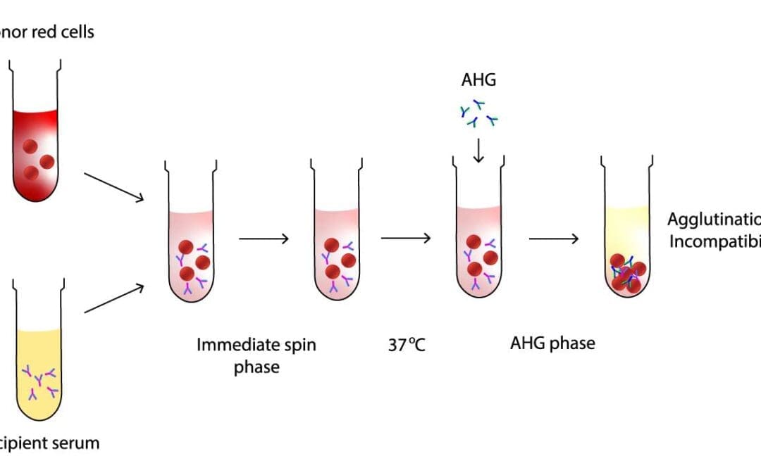
Serologic Crossmatching (Blood Compatibility Test)
Cross-matching mimics transfusion by mixing recipient serum & donor cells. Clumping (agglutination) indicates incompatibility, preventing transfusion reactions.
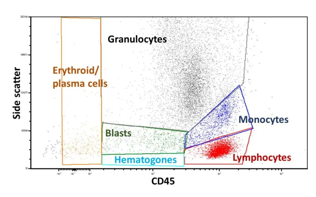
Flow Cytometry Immunophenotyping of Blood
Blood cells stained with fluorescent antibodies reveal hidden markers, like a cellular fingerprint. Flow cytometry analyzes millions of cells, painting a detailed picture of immune health, disease clues, and treatment insights.
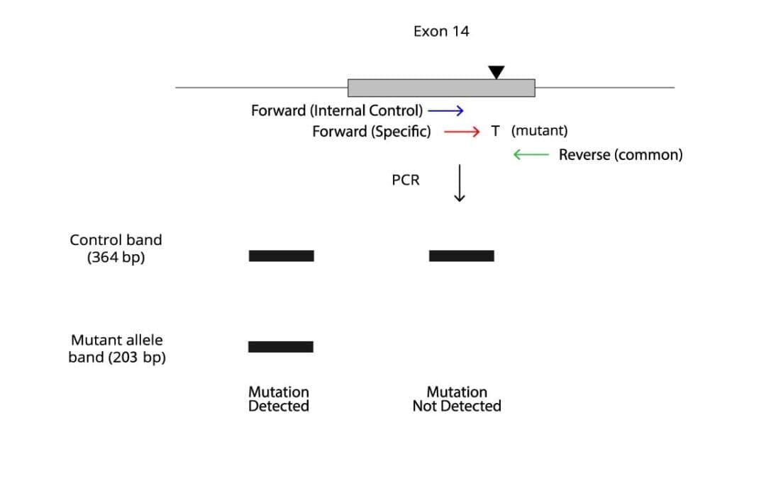
JAK2 V617F Mutation Allele Specific PCR Protocol
Uncover the JAK2 V617F mutation, a key driver in Myeloproliferative Neoplasms (MPNs). Learn ARMS PCR, a reliable technique for diagnosis!
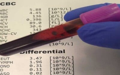
CBC with Differential and Other Reference Ranges
Reference ranges like complete blood count (CBC) with differential are important because they provide a baseline for interpreting laboratory test results.

Myeloperoxidase Reaction (MPO) Stain
A stain mainly used to differentiate AML from ALL and gives a bluish to brownish tinge in cells with lysosomal enzyme.
May-Grünwald Giemsa (MGG) Stain
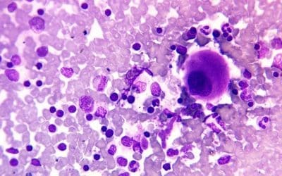
May-Grünwald Giemsa (MGG) stain is an intense Romanovsky stain to help with the visualisation of bone marrow smears.
Perls’ Prussian Blue Staining
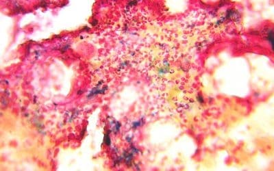
Perls’ Prussian blue stain helps in identifying presence of iron stores in the bone marrow aspirate smear.
Leishman Stain for Peripheral Blood Smear
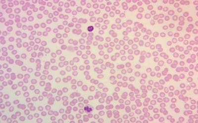
Leishman stain is used commonly for the identification of different cells present in the peripheral blood smear. It has acidic and basic properties.