Lab Protocols
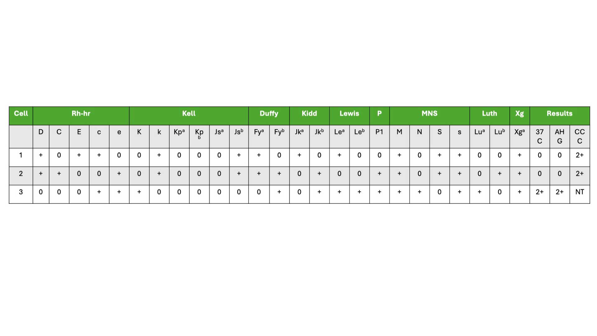
Antibody Screening
Antibody screening mixes patient plasma with red blood cells to detect unexpected antibodies. Agglutination indicates possible antibodies, requiring further identification for safe blood transfusions
Periodic Acid Schiff Stain (PAS Staining)
PAS stain is a histochemical technique that utilizes periodic acid-Schiff (PAS) reagent to detect and visualize carbohydrate-rich structures in cells and tissues, such as glycogen, glycoproteins, and mucins.
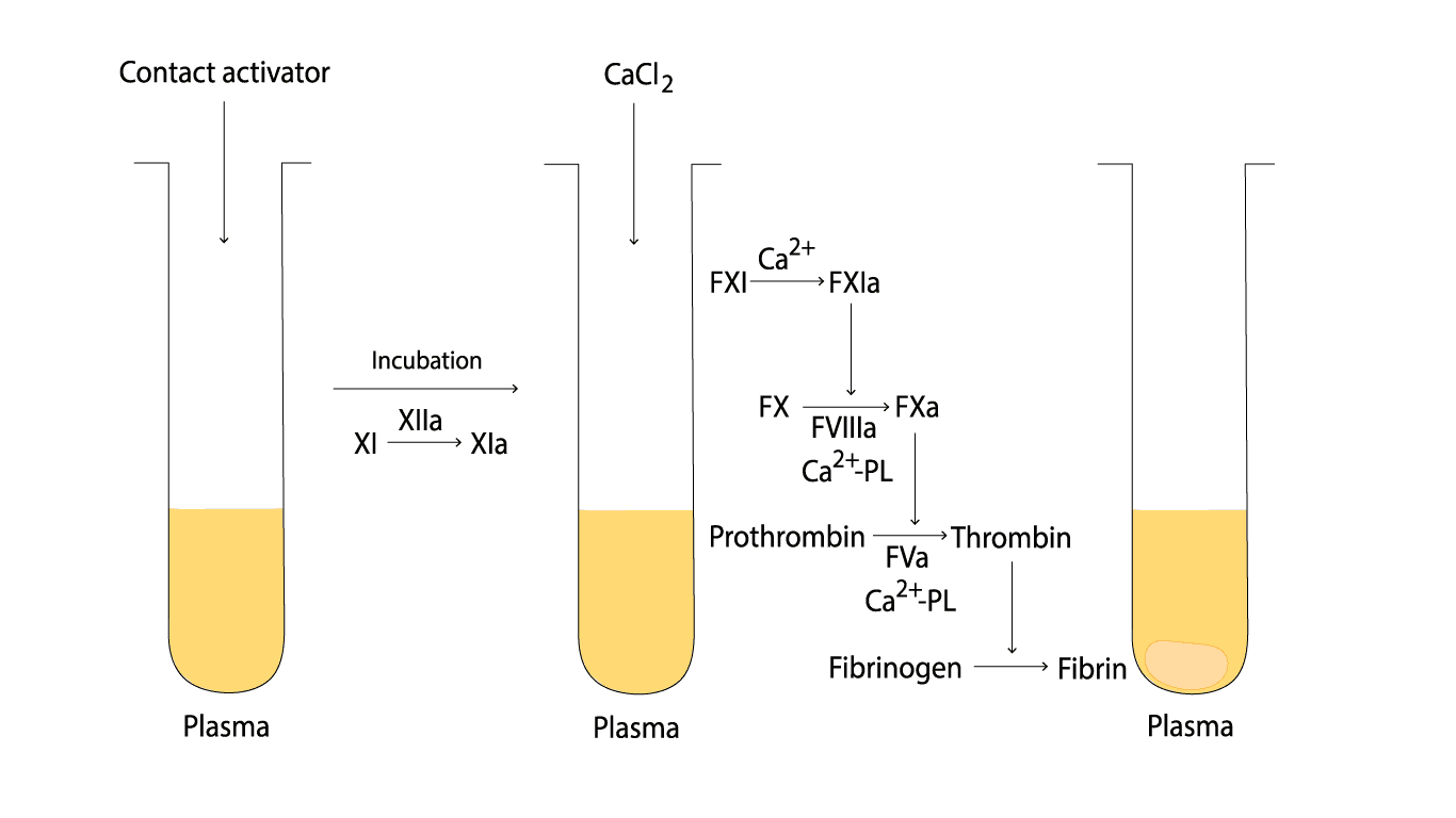
Activated Partial Thromboplastin Time (aPTT) Blood Test
The APTT assay measures how long it takes for a blood clot to form by measuring the amount of time it takes for fibrinogen to be converted to fibrin.
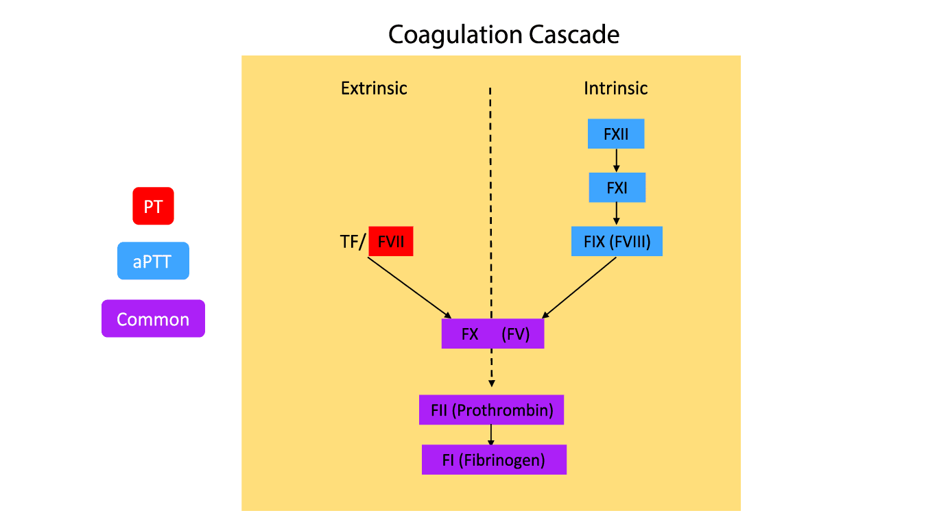
Prothrombin Time (PT) & INR
The PT assay is used to diagnose and monitor a variety of bleeding and clotting disorders by measuring how long it takes a blood clot to form.
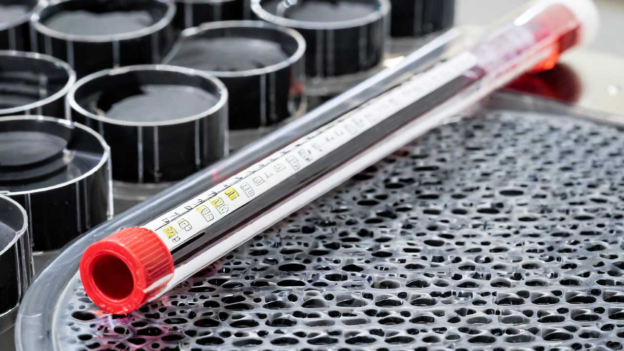
Erythrocyte Sedimentation Rate or ESR
ESR measures the sedimentation rate or the length red blood cells fall in a vertical tube over a period of time.
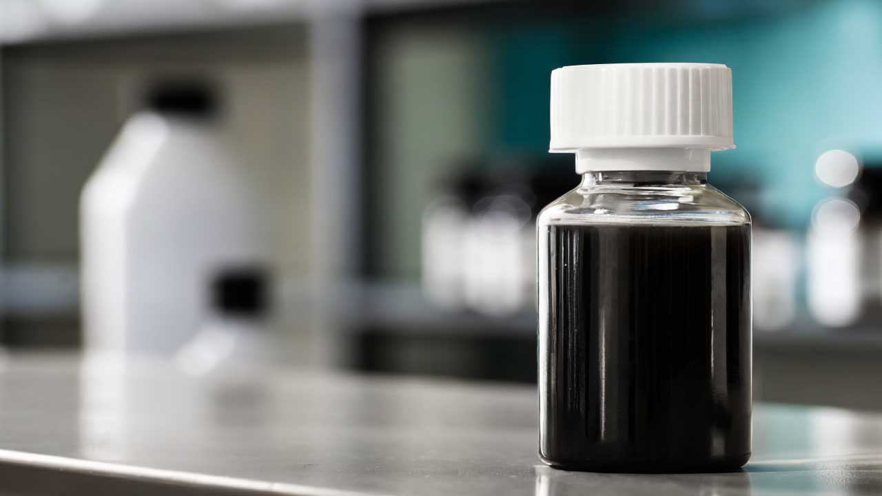
Supravital Stain: BCB or NMB
BCB or new methylene blue (NMB) stains are also known as supravital stains. These stains are commonly used to stain reticulocytes, Heinz bodies or H inclusions
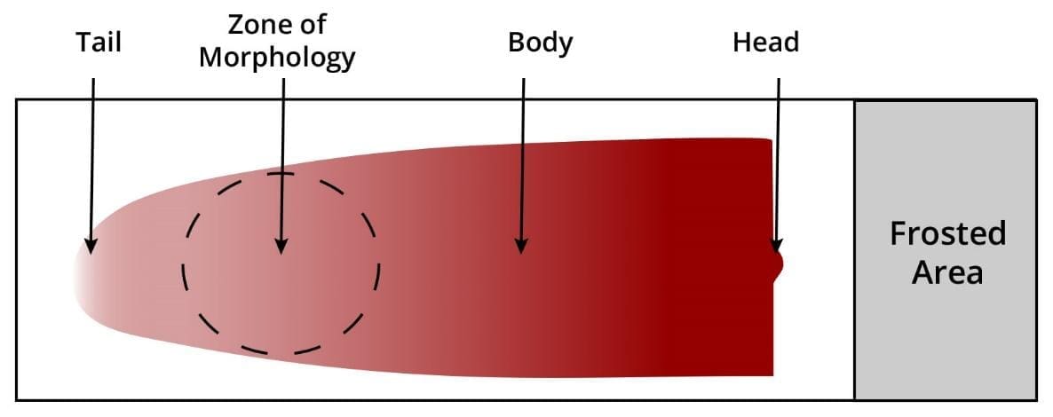
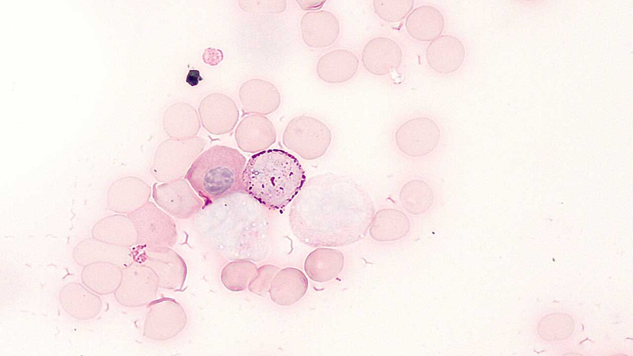
Preparation of Peripheral Blood Smears
A drop of blood spread on a slide, reveals morphological abnormalities of the blood cells that can be viewed under the microscope.