Key Takeaways
A platelet function disorder is a condition where platelets are present in normal numbers but do not stick, activate, or clump properly, so even small injuries can cause prolonged bleeding [1].
- Causes ▾: The main inherited forms are Glanzmann thrombasthenia (defective aggregation), Bernard-Soulier syndrome (defective adhesion), storage pool diseases (defective granule contents or release), and Scott syndrome (defective procoagulant surface) [4].
- Symptoms: Typical signs are mucocutaneous; easy bruising, nosebleeds, gum bleeding, heavy periods, and prolonged bleeding after minor cuts, dental work, or surgery [2].
- Diagnosis ▾: Modern diagnosis begins with the ISTH-BAT bleeding score, a full blood count, and a coagulation screen, then moves to PFA-100/PFA-200 closure times, light transmission aggregometry, flow cytometry, and selective genetic testing [1,7].
- Treatment ▾: There is no cure for most inherited forms. Management focuses on bleeding prevention, antifibrinolytics like tranexamic acid, platelet transfusions for severe events, and recombinant activated factor VII (rFVIIa) for Glanzmann patients refractory to platelets [5]. Acquired causes (antiplatelet drugs, uremia, myeloproliferative neoplasms, hyperglobulinemia) are managed by treating the underlying problem and adjusting bleeding risk [1].
*Click ▾ for more information
Introduction
Platelets stop bleeding by sticking to damaged blood vessels and clumping into a plug. When their numbers are fine but their machinery fails, the result is a platelet function disorder. A small cut may bleed for hours. A simple dental extraction can turn into a hematology emergency.
This article walks through what platelets do, what can go wrong, how the dysfunction is found in the laboratory, and how it is treated using current guidance from the International Society on Thrombosis and Haemostasis (ISTH) [1].
How Platelet Normally Work
Platelets act as the body's first responders in clotting (hemostasis).
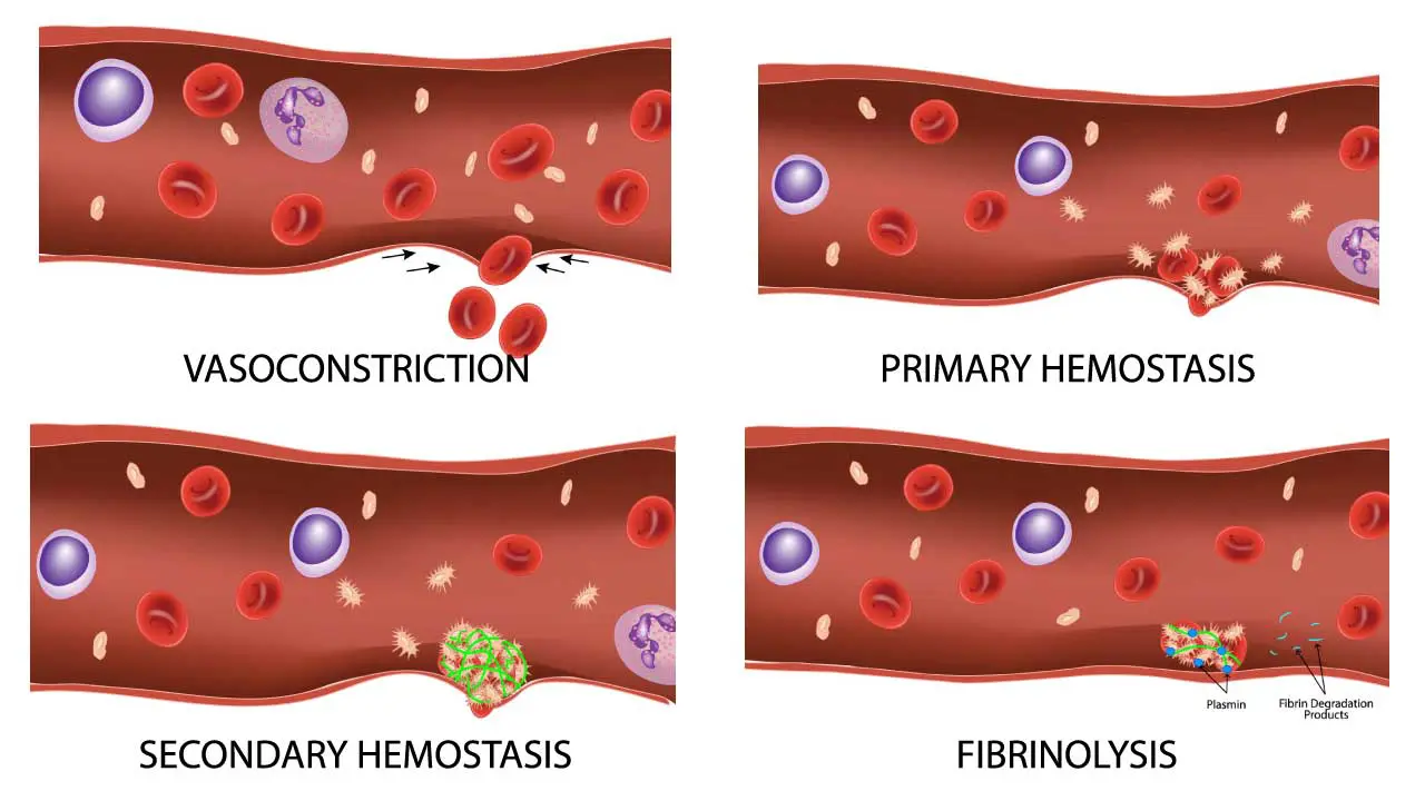
Primary hemostasis happens in four steps. Each one is a target that disease can attack.
- Adhesion. When a vessel tears, collagen is exposed. Von Willebrand factor (vWF), a plasma protein, binds collagen and acts as a bridge to platelets through the GPIb-IX-V receptor.
- Activation and shape change. Platelets become sticky, change shape, and release the contents of their internal granules (dense and alpha) [1].
- Aggregation. Activated platelets cross-link to each other using GPIIb/IIIa, which binds fibrinogen.
- Procoagulant surface. Activated platelets flip the membrane lipid phosphatidylserine to the outside. This surface lets clotting factors assemble and produce thrombin.
Each inherited platelet function disorder maps cleanly to one of these steps. Adhesion fails in Bernard-Soulier. Granule release fails in storage pool diseases. Aggregation fails in Glanzmann. The procoagulant surface fails in Scott syndrome [4]. Keeping this map in mind makes the rest of the article easier to follow.
Causes of Platelet Function Disorders
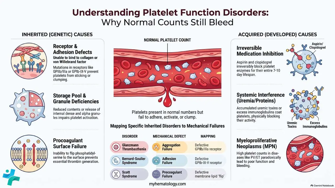
Causes fall into two groups: inherited (present from birth, genetic) and acquired (developed later in life).
Inherited Disorders
These are rare conditions caused by mutations in genes that encode platelet proteins. Most follow autosomal recessive inheritance, meaning both parents must carry the gene change for a child to be affected. A minority are autosomal dominant [1].
Glanzmann Thrombasthenia (GT)
- Defect: Mutations in ITGA2B or ITGB3 cause a deficient or non-functional GPIIb/IIIa complex. Without this receptor, platelets cannot bind fibrinogen and cannot clump together [1].
- Inheritance: Autosomal recessive.
- Symptoms: Lifelong mucocutaneous bleeding from early childhood: epistaxis (nosebleeds), gum bleeding, heavy menstrual bleeding, and bleeding after surgery or trauma. Epistaxis is the most common presenting symptom [2].
- Peripheral smear: Platelet count and size are normal.
- Diagnosis: Light transmission aggregometry shows absent aggregation with all physiological agonists (ADP, collagen, epinephrine, arachidonic acid) but normal agglutination with ristocetin. Flow cytometry confirms reduced GPIIb/IIIa expression. Genetic testing identifies the mutation [1,7].
- Treatment: Local pressure, antifibrinolytics, platelet transfusion for major bleeds, and recombinant activated factor VII (rFVIIa, NovoSeven RT) for patients refractory to platelets or with anti-platelet antibodies. rFVIIa is FDA-approved for this exact indication [5].
Bernard-Soulier Syndrome
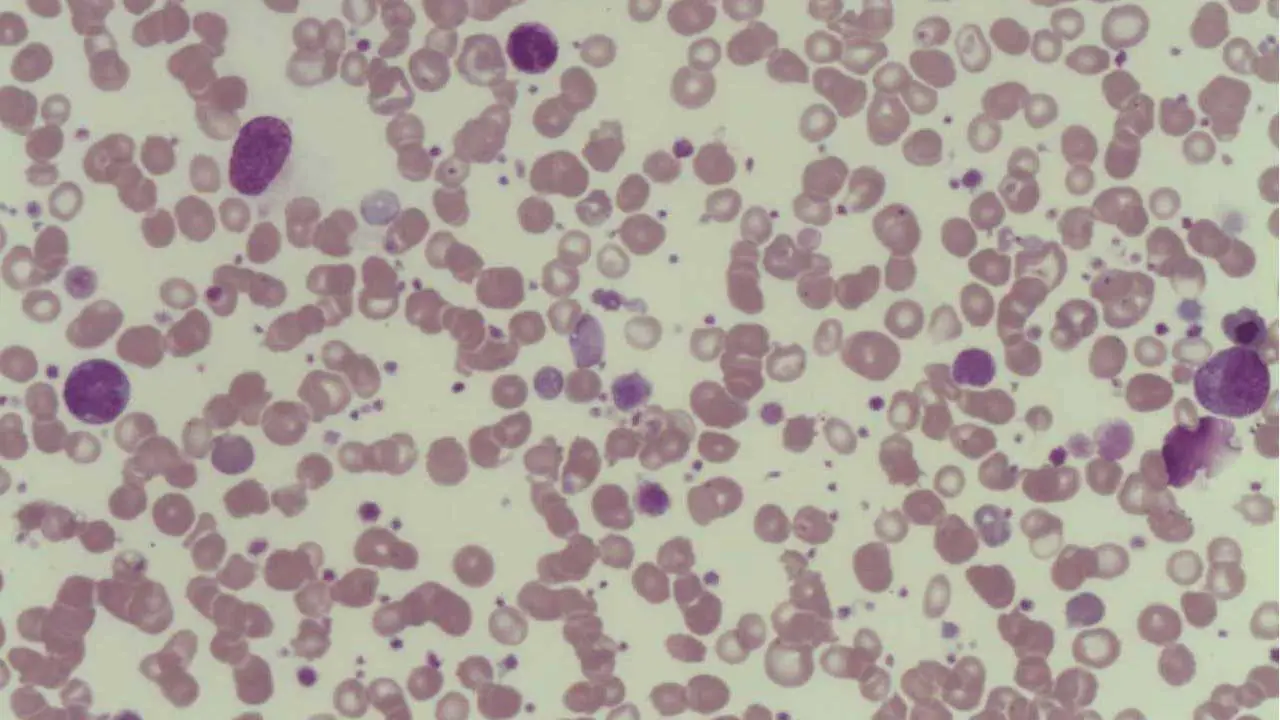
- Defect: Mutations in GP1BA, GP1BB, or GP9 cause loss of the GPIb-IX-V complex, the platelet receptor for vWF. Platelets cannot adhere to the injured vessel wall [1].
- Inheritance: Autosomal recessive (rarely dominant).
- Symptoms: Mucocutaneous bleeding similar in pattern to Glanzmann, often more severe.
- Peripheral smear: Large (giant) platelets and mild thrombocytopenia.
- Diagnosis: Aggregometry shows absent agglutination with ristocetin (does not correct with normal plasma, distinguishing it from vWF disease). Flow cytometry shows reduced GPIb expression [1,7].
- Treatment: Antifibrinolytics, platelet transfusion for major bleeds, and supportive care. Desmopressin has limited benefit.
MYH9-Related Disease (MYH9-RD)
- Defect: Mutations in the MYH9 gene cause defective non-muscle myosin heavy chain IIA, leading to impaired platelet cytoskeletal function and macrothrombocytopenia (large platelets in low numbers) [11].
- Inheritance: Autosomal dominant.
- Symptoms: Mild to moderate mucocutaneous bleeding. Patients are frequently misdiagnosed with Immune Thrombocytopenia (ITP) and may inappropriately receive corticosteroids or splenectomy before the true genetic cause is found [11].
- Peripheral smear: Giant platelets and pale blue inclusions in the cytoplasm of neutrophils (Döhle-like bodies).
- Diagnosis: Confirmed by peripheral blood smear review and MYH9 genetic testing.
- Treatment: Antifibrinolytics, desmopressin, and thrombopoietin (TPO) receptor agonists (e.g., eltrombopag) to raise platelet counts prior to major surgery [12].
Storage Pool Diseases
- Defect: Reduced contents or impaired release of platelet granules. Two main types exist [4]:
- Dense (δ) granule deficiency: lacks ADP, ATP, calcium, serotonin. Seen in syndromes like Hermansky-Pudlak and Chediak-Higashi.
- Alpha granule deficiency (Gray Platelet Syndrome): caused by NBEAL2 mutations; platelets look gray on a Wright-stained smear because alpha granule proteins are missing.
- Symptoms: Mild to moderate mucocutaneous bleeding. Some patients are diagnosed only after a bleeding challenge such as dental extraction.
- Diagnosis: Aggregometry typically shows absent secondary wave. Electron microscopy confirms granule deficiency. Flow cytometry for granule markers and genetic testing complete the workup [10].
- Treatment: Antifibrinolytics, desmopressin in selected cases, and platelet transfusion for severe bleeding.
Scott Syndrome
- Defect: Mutations in ANO6 (TMEM16F) disrupt the calcium-activated phospholipid scramblase that flips phosphatidylserine to the platelet surface. Without this procoagulant surface, thrombin generation is impaired [6].
- Inheritance: Autosomal recessive.
- Symptoms: Bleeding tendency often surfaces only after trauma or surgery, since routine aggregation tests look normal.
- Diagnosis: Annexin V binding assay shows reduced phosphatidylserine exposure after activation. Standard aggregometry is often normal, which is the diagnostic clue. Genetic testing confirms ANO6 mutation [6].
- Treatment: Antifibrinolytics and platelet transfusion when bleeding occurs.
Acquired Disorders
Acquired causes are far more common than inherited ones and are often the reason a normal-count patient bleeds after surgery.
Antiplatelet Drugs
Aspirin, clopidogrel, prasugrel, and ticagrelor are prescribed deliberately to inhibit platelet function and prevent arterial clots in heart disease and stroke. The trade-off is bleeding risk. Aspirin and clopidogrel are irreversible, so the effect lasts the lifespan of the platelet (about 7–10 days). Ticagrelor is reversible and clears faster. This matters when planning surgery [1].
Uremia
Kidney failure allows uremic toxins (such as guanidinosuccinic acid) to accumulate. These toxins impair platelet adhesion and aggregation. Bleeding usually improves with dialysis. Desmopressin, cryoprecipitate, and correction of anemia can also help [1].
Myeloproliferative Neoplasms and Myelodysplastic Syndromes
In MPNs (such as essential thrombocythemia and polycythemia vera), platelet numbers may be very high yet poorly functional. Paradoxically, patients can have both bleeding and thrombosis. In MDS, platelets are reduced in number and often dysfunctional [1].
Hyperglobulinemia
In conditions like multiple myeloma, Waldenström macroglobulinemia, lupus, and chronic infection, excess immunoglobulins coat platelets and interfere with adhesion and aggregation. Treating the underlying disease is the priority; plasma exchange can be used for urgent bleeding [1].
Other Acquired Causes
Cardiopulmonary bypass and extracorporeal membrane oxygenation (ECMO) cause transient platelet dysfunction through mechanical stress. Liver disease impairs synthesis of clotting proteins and alters platelet function [1].
Diagnosis of Platelet Function Disorders
Diagnosis is a stepwise process. The goal is to confirm dysfunction, identify the type, and rule out commoner causes like von Willebrand disease and medication effects.
Step 1: Bleeding History and ISTH-BAT
The ISTH Bleeding Assessment Tool (ISTH-BAT) is a validated questionnaire that scores lifetime bleeding episodes. A positive score in someone with a normal platelet count and normal coagulation screen is a strong trigger for further testing [2].
Step 2: Basic Laboratory Tests
A full blood count establishes platelet number and size. Coagulation tests (PT, aPTT) exclude clotting factor problems. A peripheral smear can reveal giant platelets (Bernard-Soulier) or gray platelets (Gray Platelet Syndrome).
Step 3: Screening Tests of Platelet Function
The PFA-100 / PFA-200 (Platelet Function Analyzer) is the current bench-top screen. Whole blood is drawn through a cartridge coated with collagen plus epinephrine or collagen plus ADP, and the time taken to occlude a small aperture is reported as the closure time [7]. The older bleeding time test (Template or Ivy) is no longer recommended because of poor reproducibility and weak clinical correlation; major laboratory bodies have advised removing it from routine practice [7].
However, a normal PFA-100 result does not definitively rule out a platelet function disorder. The test lacks sensitivity for mild defects, such as certain secretion defects and mild storage pool diseases. If a patient has a highly positive ISTH-BAT bleeding score but a normal PFA-100 closure time, the clinician must not stop the workup and should proceed directly to light transmission aggregometry or genetic testing [13].
Step 4: Light Transmission Aggregometry (LTA)
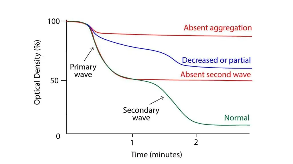
LTA remains the gold standard [3]. Platelet-rich plasma is exposed to a panel of agonists (ADP, collagen, epinephrine, arachidonic acid, ristocetin), and the change in light transmission as platelets clump is recorded. The pattern of response identifies the type of disorder. For example, absent aggregation with all agonists except ristocetin points to Glanzmann; absent ristocetin response with normal others points to Bernard-Soulier or vWF disease.
Step 5: Confirmatory Tests
- Flow cytometry quantifies surface glycoproteins (GPIIb/IIIa, GPIb-IX-V) and detects abnormal activation markers.
- Electron microscopy confirms granule deficiencies in storage pool diseases.
- Annexin V binding demonstrates the procoagulant defect of Scott syndrome.
- Genetic testing identifies the specific mutation and supports family counseling. Notably, Next-Generation Sequencing (NGS) bleeding disorder panels are increasingly used as second-line or even first-line diagnostic tools. Because traditional LTA requires large volumes of fresh blood (which is particularly challenging in pediatric patients) and highly specialized laboratory processing, many centers now bypass LTA entirely in favor of NGS panels once standard coagulation screens return normal [14].
When to Suspect a Platelet Function Disorder
A patient with mucocutaneous bleeding (skin, nose, gums, gut, menses), a normal platelet count, and normal PT and aPTT. That combination should prompt platelet function testing rather than repeating coagulation panels.
Management of Platelet Function Disorders
Management depends on whether the disorder is inherited or acquired, and on how severe the bleeding tendency is.
Inherited Disorders
There is no cure for most inherited platelet function disorders. The plan is bleeding prevention and rapid control of bleeds when they happen [1].
- Avoid aggravating drugs. Aspirin, NSAIDs, and many herbal supplements impair platelets further. Acetaminophen (paracetamol) is usually safer for pain or fever.
- Antifibrinolytics. Tranexamic acid and epsilon-aminocaproic acid prevent clots from breaking down. They are useful before dental work, during menstruation, and after minor procedures.
- Desmopressin (DDAVP). Releases stored vWF and may help in some storage pool diseases.
- Thrombopoietin (TPO) receptor agonists. Drugs such as eltrombopag or romiplostim are increasingly used off-label for inherited conditions involving functional defects combined with low platelet counts (e.g., Bernard-Soulier Syndrome and MYH9-related disease). By raising the absolute platelet count, the increased volume of platelets can often compensate for individual functional deficits, securing hemostasis for planned surgeries [12].
- Platelet transfusion. Used for serious bleeding or major surgery. Leukocyte-depleted, HLA-matched products are preferred for Glanzmann patients to reduce alloimmunization risk [5].
- Recombinant activated factor VII (rFVIIa, NovoSeven RT). FDA-approved for Glanzmann thrombasthenia refractory to platelet transfusion or with anti-platelet antibodies. Standard regimens use 90 µg/kg every 2 hours until hemostasis [5].
- Thrombosis risk awareness. While inherited platelet function disorders naturally protect against spontaneous arterial thrombosis, patients who receive multiple intensive procoagulant therapies (such as rFVIIa combined with systemic antifibrinolytics) during severe bleeds or major surgery carry a secondary, iatrogenic risk for thrombotic events. Therapy must be titrated carefully [15].
- Splenectomy. Considered in select severe cases.
- Gene therapy. Under investigation; not yet standard care.
Acquired Disorders
Treat the underlying cause whenever possible.
- Antiplatelet drugs: Balance bleeding risk against thrombosis risk. For elective surgery, individualized timing of drug discontinuation is planned with the prescribing physician.
- Uremia: Dialysis, desmopressin, cryoprecipitate, and correction of anemia.
- MPN/MDS: Disease-specific treatment (e.g., hydroxyurea, chemotherapy, stem cell transplantation in severe MDS).
- Hyperglobulinemia: Treat the underlying autoimmune, infectious, or hematologic cause; plasma exchange for severe bleeding.
Frequently Asked Questions (FAQs)
What is the difference between a platelet function disorder and thrombocytopenia?
Thrombocytopenia means the number of platelets in the blood is low. A platelet function disorder means the number is usually normal, but the platelets do not work properly. Both can cause bleeding, but the tests used to find them are different.
How to diagnose a platelet function disorder?
Diagnosis usually starts with a bleeding history using the ISTH-BAT questionnaire and a full blood count to confirm a normal platelet number. Modern screens include the PFA-100 or PFA-200. Confirmation comes from light transmission aggregometry, flow cytometry for platelet surface receptors, and, in selected cases, genetic testing. Old-style bleeding time tests are no longer recommended.
Can platelet function disorders be cured?
Most inherited forms cannot be cured at present. Treatment focuses on preventing bleeding, controlling bleeds when they happen, and avoiding drugs that make platelets work even less. Gene therapy is being studied but is not yet standard care.
Why are aspirin and ibuprofen a problem for people with platelet function disorders?
Aspirin and other NSAIDs block a platelet enzyme called cyclooxygenase-1, which reduces platelet activation. In a healthy person this is mild. In someone whose platelets are already defective, it can trigger serious bleeding. Acetaminophen (paracetamol) is generally a safer choice for pain or fever.
What should caregivers do before a child with Glanzmann thrombasthenia has dental work or surgery?
The dental or surgical team should be told well in advance. Planning usually includes a hematology consultation, antifibrinolytic drugs like tranexamic acid, and, when needed, platelet transfusion or recombinant factor VIIa. Routine NSAIDs should be stopped, and the child should wear a medical alert identifier.
Are inherited platelet function disorders passed to children?
Many follow autosomal recessive inheritance, meaning both parents must carry the gene change for a child to be affected. Some, like certain forms of storage pool disease, can be autosomal dominant. Genetic counseling helps families understand recurrence risk and testing options.
Glossary of Related Medical Terms
- Platelet function disorder (platelet dysfunction): A condition in which platelets are present in normal numbers but do not work properly to stop bleeding.
- Primary hemostasis: The first phase of bleeding control, in which platelets stick to a damaged vessel wall and clump together to form a temporary plug.
- Aggregation: Platelets sticking to each other to build a plug.
- Adhesion: Platelets sticking to the damaged vessel wall, mainly through von Willebrand factor and the GPIb-IX-V receptor.
- Glycoprotein (GP) IIb/IIIa: The platelet receptor that binds fibrinogen and lets platelets stick to each other. Defective in Glanzmann thrombasthenia.
- Glycoprotein (GP) Ib-IX-V: The platelet receptor for von Willebrand factor. Defective in Bernard-Soulier syndrome.
- Von Willebrand factor (vWF): A plasma protein that acts as the bridge between platelets and an injured vessel wall.
- Granules (dense and alpha): Storage packets inside platelets. Dense granules hold ADP, ATP, calcium, and serotonin. Alpha granules hold proteins like fibrinogen and vWF.
- Storage pool disease: A platelet disorder caused by reduced contents of dense or alpha granules.
- Phosphatidylserine: A membrane lipid that platelets flip to their outer surface during activation to support clotting reactions. Defective exposure causes Scott syndrome.
- Antifibrinolytic: A drug, such as tranexamic acid, that stops clots from breaking down too early.
- Desmopressin (DDAVP): A synthetic hormone that boosts release of vWF and some platelet contents from storage.
- Recombinant activated factor VII (rFVIIa, NovoSeven): A bypass clotting agent used to stop bleeding in Glanzmann patients who no longer respond to platelet transfusions.
- Uremia: Buildup of waste products in the blood from kidney failure, which can impair platelet function.
- ISTH-BAT: A standardized questionnaire that scores a patient's lifetime bleeding history.
- PFA-100/PFA-200: A bench-top instrument that measures how quickly platelets close a small hole in a coated membrane under flow. A modern screen for platelet function.
- Light transmission aggregometry (LTA): The gold-standard laboratory test for platelet function, measuring how much light passes through plasma as platelets clump.
Disclaimer: This article is intended for educational and informational purposes only. It is not intended to be a substitute for informed professional medical advice, diagnosis, or treatment. While the information presented here is derived from credible medical sources and is believed to be accurate and up-to-date, it is not guaranteed to be complete or error-free. See additional information.
References
- Gresele, P., & Subcommittee on Platelet Physiology of the International Society on Thrombosis and Hemostasis (2015). Diagnosis of inherited platelet function disorders: guidance from the SSC of the ISTH. Journal of thrombosis and haemostasis : JTH, 13(2), 314–322. https://doi.org/10.1111/jth.12792
- Gresele, P., Orsini, S., Noris, P., Falcinelli, E., Alessi, M. C., Bury, L., Borhany, M., Santoro, C., Glembotsky, A. C., Cid, A. R., Tosetto, A., De Candia, E., Fontana, P., Guglielmini, G., Pecci, A., & BAT-VAL study investigators (2020). Validation of the ISTH/SSC bleeding assessment tool for inherited platelet disorders: A communication from the Platelet Physiology SSC. Journal of thrombosis and haemostasis : JTH, 18(3), 732–739. https://doi.org/10.1111/jth.14683
- Cattaneo, M., Cerletti, C., Harrison, P., Hayward, C. P., Kenny, D., Nugent, D., Nurden, P., Rao, A. K., Schmaier, A. H., Watson, S. P., Lussana, F., Pugliano, M. T., & Michelson, A. D. (2013). Recommendations for the Standardization of Light Transmission Aggregometry: A Consensus of the Working Party from the Platelet Physiology Subcommittee of SSC/ISTH. Journal of thrombosis and haemostasis : JTH, 10.1111/jth.12231. Advance online publication. https://doi.org/10.1111/jth.12231
- Nurden, A. T., & Nurden, P. (2015). Inherited disorders of platelet function: selected updates. Journal of thrombosis and haemostasis : JTH, 13 Suppl 1, S2–S9. https://doi.org/10.1111/jth.12898
- Poon M-C, d’Oiron R, Zotz RB, Bindslev N, Di Minno MND, Di Minno G. The international, prospective Glanzmann Thrombasthenia Registry: treatment and outcomes in surgical intervention. Haematologica 2015; 100(8):1038-1044; https://doi.org/10.3324/haematol.2014.121384.
- Suzuki, J., Umeda, M., Sims, P. J., & Nagata, S. (2010). Calcium-dependent phospholipid scrambling by TMEM16F. Nature, 468(7325), 834–838. https://doi.org/10.1038/nature09583
- Harrison, P., & Lordkipanidzé, M. (2013). Testing platelet function. Hematology/oncology clinics of North America, 27(3), 411–441. https://doi.org/10.1016/j.hoc.2013.03.003
- Hayward, C. P., Moffat, K. A., & Liu, Y. (2012). Laboratory investigations for bleeding disorders. Seminars in thrombosis and hemostasis, 38(7), 742–752. https://doi.org/10.1055/s-0032-1326780
- Peterson, W., Martin, R., Arnold, D., Carvalho, B., Cuker, A., Gadsden, J., Provan, D., Rydz, N., Shore, E., Kuter, D., Kouides, P., Lavin, M., James, P., Engen, D., & Sholzberg, M. (2025). Delphi consensus recommendations for neuraxial anesthesia in adults with platelet disorders and coagulation defects: communication from the ISTH SSC Subcommittee on von Willebrand Factor. Journal of thrombosis and haemostasis : JTH, 23(6), 2039–2049. https://doi.org/10.1016/j.jtha.2025.01.019
- Mezzano, D., Harrison, P., Frelinger, A. L., 3rd, Mumford, A. D., Noris, P., Lordkipanidzé, M., & Gresele, P. (2022). Expert opinion on the use of platelet secretion assay for the diagnosis of inherited platelet function disorders: Communication from the ISTH SSC Subcommittee on Platelet Physiology. Journal of thrombosis and haemostasis : JTH, 20(9), 2127–2135. https://doi.org/10.1111/jth.15781
- Pecci, A., Klersy, C., Gresele, P., Lee, K.J.D., De Rocco, D., Bozzi, V., Russo, G., Heller, P.G., Loffredo, G., Ballmaier, M., Fabris, F., Beggiato, E., Kahr, W.H.A., Pujol-Moix, N., Platokouki, H., Van Geet, C., Noris, P., Yerram, P., Hermans, C., Gerber, B., Economou, M., De Groot, M., Zieger, B., De Candia, E., Fraticelli, V., Kersseboom, R., Piccoli, G.B., Zimmermann, S., Fierro, T., Glembotsky, A.C., Vianello, F., Zaninetti, C., Nicchia, E., Güthner, C., Baronci, C., Seri, M., Knight, P.J., Balduini, C.L. and Savoia, A. (2014), MYH9-Related Disease: A Novel Prognostic Model to Predict the Clinical Evolution of the Disease Based on Genotype–Phenotype Correlations. Human Mutation, 35: 236-247. https://doi.org/10.1002/humu.22476
- Zaninetti, C., Gresele, P., Bertomoro, A., Klersy, C., De Candia, E., Veneri, D., Barozzi, S., Fierro, T., Alberelli, M. A., Musella, V., Noris, P., Fabris, F., Balduini, C. L., & Pecci, A. (2020). Eltrombopag for the treatment of inherited thrombocytopenias: a phase II clinical trial. Haematologica, 105(3), 820–828. https://doi.org/10.3324/haematol.2019.223966
- Quiroga, T., Goycoolea, M., Muñoz, B., Morales, M., Aranda, E., Panes, O., Pereira, J., & Mezzano, D. (2004). Template bleeding time and PFA-100 have low sensitivity to screen patients with hereditary mucocutaneous hemorrhages: comparative study in 148 patients. Journal of thrombosis and haemostasis : JTH, 2(6), 892–898. https://doi.org/10.1111/j.1538-7836.2004.00693.x
- Downes, K., Megy, K., Duarte, D., Vries, M., Gebhart, J., Hofer, S., Shamardina, O., Deevi, S. V. V., Stephens, J., Mapeta, R., Tuna, S., Al Hasso, N., Besser, M. W., Cooper, N., Daugherty, L., Gleadall, N., Greene, D., Haimel, M., Martin, H., Papadia, S., … Freson, K. (2019). Diagnostic high-throughput sequencing of 2396 patients with bleeding, thrombotic, and platelet disorders. Blood, 134(23), 2082–2091. https://doi.org/10.1182/blood.2018891192
- Di Minno, G., Zotz, R. B., d'Oiron, R., Bindslev, N., Di Minno, M. N., Poon, M. C., & Glanzmann Thrombasthenia Registry Investigators (2015). The international, prospective Glanzmann Thrombasthenia Registry: treatment modalities and outcomes of non-surgical bleeding episodes in patients with Glanzmann thrombasthenia. Haematologica, 100(8), 1031–1037. https://doi.org/10.3324/haematol.2014.121475