Key Takeaways
Pernicious anemia is an autoimmune disease in which the immune system destroys the stomach's parietal cells, blocking the production of intrinsic factor and causing vitamin B12 deficiency.
- Symptoms ▾: Typical features include macrocytic anemia, fatigue, glossitis, and neurological signs ranging from tingling in the hands and feet to subacute combined degeneration of the spinal cord.
- Diagnosis ▾: Diagnosis rests on a full blood count showing macrocytosis, a low serum vitamin B12, and positive intrinsic factor antibodies. Methylmalonic acid and homocysteine support borderline cases.
- Treatment ▾: Treatment is lifelong vitamin B12 replacement, most often intramuscular hydroxocobalamin. High-dose oral cyanocobalamin is now a recognized alternative for selected patients [2,6].
*Click ▾ for more information
Introduction
Pernicious anemia links three systems in one story: the immune system, the stomach, and the bone marrow. The disease begins when the body attacks its own parietal cells, the stomach cells that make intrinsic factor (a protein needed to absorb vitamin B12). Without intrinsic factor, B12 cannot pass from the gut into the blood. Over time the body's stores run out, red blood cell production falters, and nerves begin to suffer.
Pathogenesis
Intrinsic factor and absorption
Vitamin B12, also called cobalamin, is essential for DNA synthesis in dividing cells and for the myelin sheath that insulates nerves. The body cannot make it, so it must come from animal-based foods.
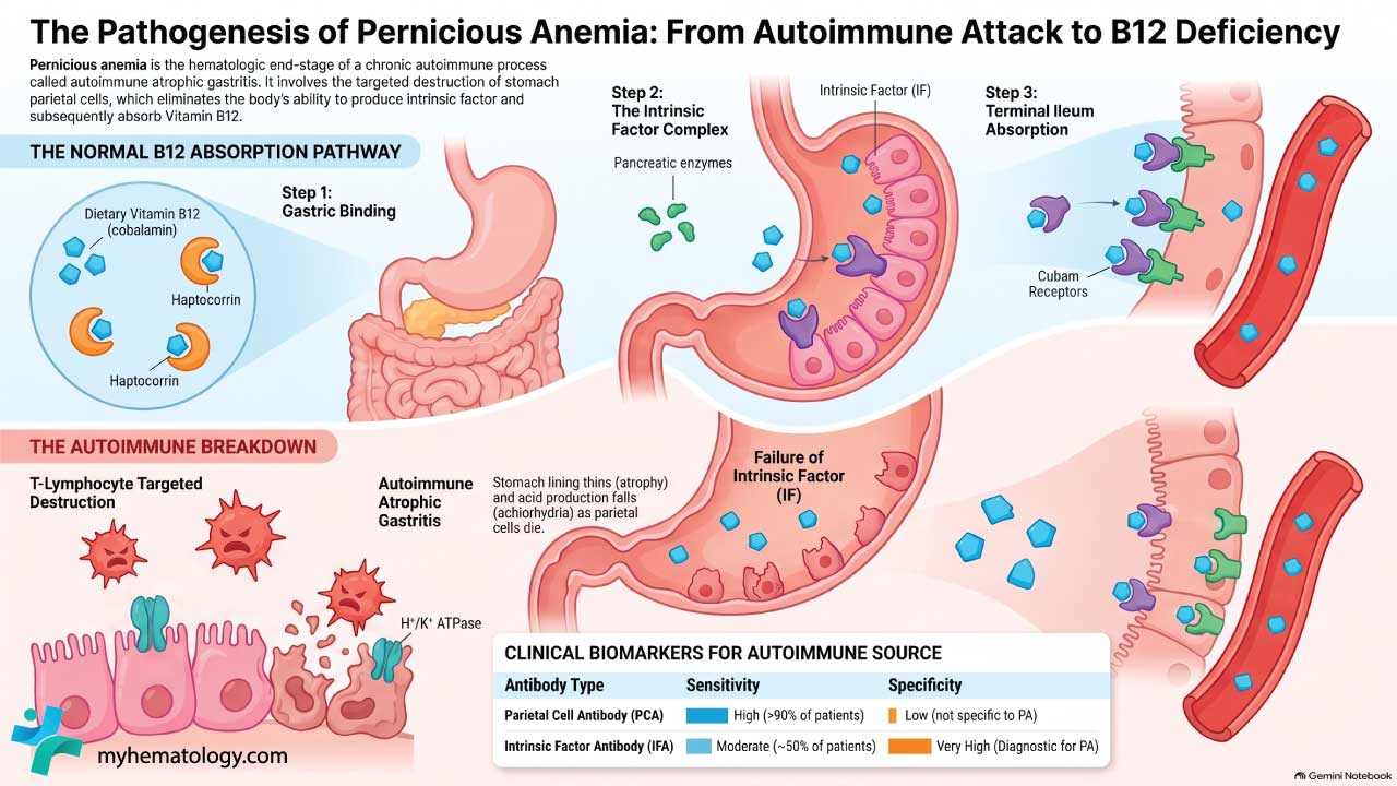
Absorption is not simple. Dietary B12 first binds to a protein called haptocorrin in the stomach. In the duodenum, pancreatic enzymes release the B12, which then binds to intrinsic factor produced by parietal cells. The intrinsic factor-B12 complex travels to the terminal ileum, the last part of the small intestine, where it docks with the cubam receptor and is absorbed into the blood. Remove any step and absorption collapses [5].
Autoimmune atrophic gastritis: the underlying lesion
Pernicious anemia does not appear out of nowhere. It is the end stage of a slower process called autoimmune atrophic gastritis, in which T lymphocytes target the H+/K+ ATPase enzyme on parietal cells. Over years, parietal cells are destroyed, the gastric body and fundus thin out (atrophy), and stomach acid production falls (achlorhydria). When parietal cell loss is severe enough, intrinsic factor production fails and B12 deficiency follows [3,7].
Two autoantibodies appear in this process and are useful clinically:
- Parietal cell antibody (PCA) is found in more than 90% of patients but is not very specific.
- Intrinsic factor antibody (IFA) is found in around half of patients and is highly specific. A positive IFA strongly supports the diagnosis [1].
The 2024 NICE guideline on vitamin B12 deficiency has prompted a wider debate about whether "autoimmune gastritis" should replace "pernicious anemia" as the preferred term, since pernicious anemia is really the hematologic complication of a longer gastric disease [2,7]. For now, both terms are in use.
Pernicious Anemia Symptoms and Signs

Symptoms develop slowly, often over years. Many people compensate for a long time before the picture becomes obvious. Three groups of symptoms are worth knowing.
Blood-related symptoms
The anemia itself produces fatigue, breathlessness on exertion, pale skin, and palpitations. These are the symptoms of any chronic anemia and are not specific to pernicious anemia.
Neurological symptoms
This is where pernicious anemia separates itself from other anemias. B12 is needed to maintain myelin, and deficiency damages peripheral nerves and the spinal cord.
- Paresthesias (tingling and numbness) usually start symmetrically in the feet and hands.
- Subacute combined degeneration of the spinal cord is the classic syndrome. It affects the dorsal columns (causing loss of vibration and position sense) and the lateral corticospinal tracts (causing weakness and spasticity). Patients lose balance, particularly with eyes closed (a positive Romberg sign).
- Cognitive changes range from poor concentration and low mood to frank dementia in severe, untreated cases.
Neurological symptoms can occur without anemia. A normal hemoglobin does not rule out B12 deficiency [1,5].
Gastrointestinal symptoms
These are often subtle or absent.
- Glossitis, a smooth, red, sore tongue, is a classic sign.
- Diarrhea or mild dyspepsia may occur because of atrophic gastritis.

Pernicious Anemia Laboratory Investigations
Diagnosis of pernicious anemia combines a hematologic picture with biochemical confirmation of B12 deficiency and serological evidence of autoimmunity.

Complete blood count and peripheral smear
- Macrocytosis (raised MCV, often >100 fL) is the hallmark. Pernicious anemia is one of the megaloblastic anemias.
- Hypersegmented neutrophils on the blood smear (five or more lobes) are a classic, early finding.
- Pancytopenia can occur in severe deficiency because megaloblastic change affects all three cell lines.
- Co-existing iron deficiency is common because achlorhydria impairs iron absorption. When both deficiencies are present, the picture is dimorphic (a mixed population of large and small cells), and the MCV may sit in the normal range.
Vitamin B12 and metabolic markers
- Serum B12 below the laboratory reference range supports the diagnosis, but borderline values are common and can be misleading.
- Methylmalonic acid (MMA) and homocysteine rise in true B12 deficiency at the tissue level and help clarify equivocal cases.
- Holotranscobalamin (active B12) measures only the metabolically usable fraction of B12 and is increasingly used as a more sensitive first-line test [2,5].
Current guidelines emphasize treating the patient rather than relying solely on the laboratory test. If a patient presents with strong clinical features of B12 deficiency especially neurological symptoms, treatment should be initiated even if total serum B12 levels fall within the normal or borderline range [2]. Furthermore, B12 testing must always be paired with serum folate (Vitamin B9). If a patient is deficient in both, vitamin B12 must be replaced first. Administering folate to a B12-deficient patient will drive DNA synthesis and correct the macrocytic anemia, but it will rapidly consume the remaining trace B12 stores. This precipitates or accelerates irreversible subacute combined degeneration of the spinal cord [1,5].
Autoantibody and gastric workup
- Intrinsic factor antibody (IFA): highly specific; a positive result is often considered diagnostic in the right clinical context.
- Parietal cell antibody (PCA): sensitive but not specific.
- Gastrin is typically elevated, reflecting loss of acid feedback.
- Endoscopy with biopsy is recommended at diagnosis to confirm atrophic gastritis, stage it (using the OLGA or OLGIM systems), and exclude prevalent gastric neoplasia [3,9].
- The Schilling test is largely obsolete and is no longer performed in most centers.
Pernicious Anemia Treatment and Management
Pernicious anemia is highly treatable. The aim is to restore vitamin B12 stores, reverse symptoms, and prevent long-term complications, especially the spinal cord damage that gives the disease its name.
Vitamin B12 replacement: the cornerstone
In the UK and much of Europe, hydroxocobalamin is the parenteral form of choice because it binds plasma proteins tightly and gives a longer duration of action. In the United States, cyanocobalamin is more commonly used.
A typical regimen for pernicious anemia is:
- Loading: 1 mg of hydroxocobalamin intramuscularly on alternate days for 2 weeks, or until no further improvement is seen if neurological symptoms are present.
- Maintenance: 1 mg intramuscularly every 2–3 months for life [1,2].
It is vital to note that if any neurological symptoms are present, intramuscular replacement is strictly mandated to ensure rapid and guaranteed tissue delivery; oral therapy is heavily contraindicated for the acute reversal of neurological deficits [1,2].
Oral vitamin B12: a valid alternative
About 1% of an oral dose of B12 is absorbed by passive diffusion, independent of intrinsic factor. At doses of 1,000–2,000 micrograms daily, this is enough to correct deficiency in many patients with pernicious anemia. Randomized trials have shown comparable improvement in serum B12, MMA, and hematologic markers between high-dose oral and intramuscular regimens [6]. Current guidelines, including NICE 2024, accept oral replacement as an option, particularly for maintenance in patients without active neurological involvement [2]. Patient preference, adherence, and access matter. Some patients report better symptom control with injections; others prefer the convenience of a daily tablet.
Monitoring response
A reticulocyte rise within a week, followed by normalization of the MCV over 6–8 weeks, confirms a clinical response. However, this sudden burst of bone marrow activity carries acute metabolic risks. The rapid production of new red blood cells pulls massive amounts of potassium into the intracellular space, which can precipitate sudden and severe hypokalemia.
Serum potassium must be closely monitored during the first 48 hours of B12 therapy in patients presenting with severe anemia [1]. Similarly, this surge in hematopoiesis will aggressively consume iron stores. Even if iron levels are normal at baseline, iron depletion often becomes apparent once megaloblastic suppression is reversed, and caregivers should anticipate the need for iron supplementation in the weeks following initial B12 loading [1,5].
Managing neurological complications
Early B12 replacement reverses most peripheral symptoms. Long-standing subacute combined degeneration may improve only partially. Physical and occupational therapy support recovery, and neuropathic pain medications can be used for persistent paresthesia.
Gastric cancer surveillance
Patients with pernicious anemia have an estimated 2- to 7-fold increased risk of gastric adenocarcinoma and a markedly increased risk of Type 1 gastric carcinoid (neuroendocrine) tumors [8]. The risk is driven by the underlying atrophic gastritis, not by B12 deficiency itself. Current AGA and European MAPS II guidance recommend:
- A baseline endoscopy with mapped biopsies at diagnosis.
- Surveillance endoscopy every 3 years for patients with extensive atrophy or intestinal metaplasia (OLGA/OLGIM stage III–IV) [3,9].
Associated autoimmune conditions
Pernicious anemia clusters with other organ-specific autoimmune diseases. The most commonly associated are autoimmune thyroid disease (Hashimoto thyroiditis, Graves disease), Type 1 diabetes mellitus, Addison disease, vitiligo, and celiac disease. Screening thresholds vary, but thyroid function tests are reasonable at diagnosis. Celiac disease is worth considering when iron deficiency or chronic gastrointestinal symptoms persist despite B12 replacement.
Prognosis
With early diagnosis and consistent treatment, the outlook for pernicious anemia is very good. Anemia and most peripheral neurological symptoms resolve within weeks to months. Severe or long-standing spinal cord damage may not fully reverse, which is why prompt recognition matters so much. Lifelong B12 replacement and periodic gastric surveillance allow most patients to live without significant limitation [1,2].
Three factors influence outcome:
- Duration of deficiency before diagnosis. The longer the delay, the higher the chance of irreversible neurological damage.
- Adherence to replacement therapy. Whether injections or oral, consistency matters more than the route.
- Management of co-existing conditions, especially iron deficiency, thyroid disease, and atrophic gastritis surveillance.
Differential Diagnosis
- Dietary deficiency in strict vegans, particularly during pregnancy and infancy.
- Medications: metformin and long-term proton pump inhibitors reduce B12 absorption.
- Ileal disease or resection: Crohn disease, ileal Crohn surgery, or extensive small bowel disease impair the final absorption step.
- Bacterial overgrowth in the small intestine consumes B12 before it reaches the ileum.
- Helicobacter pylori gastritis can contribute to both atrophic gastritis and B12 malabsorption and is often tested for and eradicated when present.
The key distinguishing features of pernicious anemia are positive intrinsic factor antibodies and biopsy evidence of autoimmune atrophic gastritis.
Why This Matters
Pernicious anemia is one of the few neurological emergencies in hematology that is fully treatable with a simple injection. A delayed diagnosis can leave a patient with permanent spinal cord damage. An early one restores them to full function.
Frequently Asked Questions (FAQs)
What causes pernicious anemia?
Pernicious anemia is caused by an autoimmune attack on the parietal cells of the stomach lining. These cells normally make intrinsic factor, a protein that vitamin B12 needs in order to be absorbed in the small intestine. Without intrinsic factor, B12 absorption fails and a deficiency develops, even when dietary intake is adequate.
How is pernicious anemia diagnosed?
Diagnosis usually starts with a complete blood count showing enlarged red blood cells (macrocytic anemia) and a low serum vitamin B12 level. The diagnosis is confirmed by testing for intrinsic factor antibodies and, in some cases, parietal cell antibodies. Methylmalonic acid and homocysteine can confirm tissue-level deficiency when serum B12 is borderline. Endoscopy with biopsy may be used to confirm the underlying autoimmune atrophic gastritis.
What is the difference between pernicious anemia and vitamin B12 deficiency?
Vitamin B12 deficiency is the broad term for any cause of low B12, including poor diet, gut disease, certain medications (metformin, proton pump inhibitors), or surgery. Pernicious anemia is one specific cause, defined by the autoimmune destruction of parietal cells and loss of intrinsic factor.
How is pernicious anemia treated?
The standard treatment is vitamin B12 replacement. In most cases this means intramuscular hydroxocobalamin, given as a loading course (often 1 mg on alternate days for two weeks) followed by 1 mg every 2 to 3 months for life. High-dose oral cyanocobalamin (1,000–2,000 micrograms daily) is an effective alternative for many patients without active neurological symptoms.
Is pernicious anemia linked to stomach cancer?
Yes. Pernicious anemia carries an increased risk of gastric adenocarcinoma and Type 1 gastric carcinoid tumors. The risk traces to the underlying atrophic gastritis, not to B12 deficiency itself. Current guidelines recommend a baseline endoscopy at diagnosis, with surveillance every three years in patients who have extensive atrophy on biopsy.
Can the nerve damage from pernicious anemia be reversed?
Some neurological symptoms reverse fully with prompt B12 replacement, especially early paresthesias and cognitive fog. However, if treatment is delayed for months or years, damage to the spinal cord (subacute combined degeneration) may be only partially reversible. Early recognition and treatment are the strongest predictors of full recovery.
Glossary of Related Medical Terms
- Pernicious anemia: An autoimmune disease in which the immune system attacks stomach cells that make intrinsic factor, causing vitamin B12 deficiency.
- Intrinsic factor: A protein made by stomach parietal cells. It binds vitamin B12 so the body can absorb it in the lower small intestine.
- Parietal cell: A specialized cell in the stomach lining that makes stomach acid and intrinsic factor.
- Vitamin B12 (cobalamin): A water-soluble vitamin essential for making red blood cells and for healthy nerve function.
- Autoimmune atrophic gastritis: Chronic inflammation and thinning of the stomach lining caused by an immune attack on parietal cells. It is the underlying lesion in pernicious anemia.
- Macrocytosis: Larger-than-normal red blood cells, measured by mean corpuscular volume (MCV) above the reference range.
- Megaloblastic anemia: A type of anemia in which red blood cell precursors in the bone marrow are abnormally large because of impaired DNA synthesis, usually from B12 or folate deficiency.
- Methylmalonic acid (MMA): A metabolite that builds up when vitamin B12 is low. A sensitive marker of true B12 deficiency.
- Homocysteine: An amino acid that rises when either B12 or folate is deficient.
- Holotranscobalamin (active B12): The fraction of vitamin B12 bound to transcobalamin and available for cellular use. An early marker of deficiency.
- Subacute combined degeneration: Damage to the dorsal columns and lateral corticospinal tracts of the spinal cord caused by B12 deficiency. Produces numbness, weakness, and balance problems.
- Achlorhydria: Absence of stomach acid. A consequence of parietal cell loss.
- Hydroxocobalamin: The form of vitamin B12 most commonly used for injection in the UK and Europe.
- Cyanocobalamin: A synthetic form of vitamin B12 used in oral supplements and, in some countries, for injection.
- Schilling test: An older radioactive test of B12 absorption, now largely obsolete.
Disclaimer: This article is intended for educational and informational purposes only. It is not intended to be a substitute for informed professional medical advice, diagnosis, or treatment. While the information presented here is derived from credible medical sources and is believed to be accurate and up-to-date, it is not guaranteed to be complete or error-free. See additional information.
References
- Devalia, V., Hamilton, M. S., Molloy, A. M., & British Committee for Standards in Haematology (2014). Guidelines for the diagnosis and treatment of cobalamin and folate disorders. British journal of haematology, 166(4), 496–513. https://doi.org/10.1111/bjh.12959
- National Institute for Health and Care Excellence. (2024). Vitamin B12 deficiency in over 16s: Diagnosis and management (NICE Guideline NG239).
- Shah, S. C., Piazuelo, M. B., Kuipers, E. J., & Li, D. (2021). AGA Clinical Practice Update on the Diagnosis and Management of Atrophic Gastritis: Expert Review. Gastroenterology, 161(4), 1325–1332.e7. https://doi.org/10.1053/j.gastro.2021.06.078
- Patel, H., & McGuirk, R. (2025). Vitamin B12 Deficiency: Common Questions and Answers. American family physician, 112(3), 294–300.
- Wolffenbuttel, B. H., Owen, P. J., Ward, M., & Green, R. (2023). Vitamin B12. BMJ (Clinical research ed.), 383, e071725. https://doi.org/10.1136/bmj-2022-071725
- Sanz-Cuesta, T., Escortell-Mayor, E., Cura-Gonzalez, I., Martin-Fernandez, J., Riesgo-Fuertes, R., Garrido-Elustondo, S., Mariño-Suárez, J. E., Álvarez-Villalba, M., Gómez-Gascón, T., González-García, I., González-Escobar, P., Vargas-Machuca Cabañero, C., Noguerol-Álvarez, M., García de Blas-González, F., Baños-Morras, R., Díaz-Laso, C., Caballero-Ramírez, N., Herrero de-Dios, A., Fernández-García, R., Herrero-Hernández, J., … OB12 Group (2020). Oral versus intramuscular administration of vitamin B12 for vitamin B12 deficiency in primary care: a pragmatic, randomised, non-inferiority clinical trial (OB12). BMJ open, 10(8), e033687. https://doi.org/10.1136/bmjopen-2019-033687
- Thain, A., Hart, K., & Ahmadi, K. R. (2025). Addressing the Gaps in the Vitamin B12 Deficiency 2024 NICE Guidelines: Highlighting the Need for Better Recognition, Diagnosis, and Management of Pernicious Anaemia. European journal of clinical nutrition, 79(7), 607–610. https://doi.org/10.1038/s41430-025-01583-4
- Murphy, G., Dawsey, S. M., Engels, E. A., Ricker, W., Parsons, R., Etemadi, A., Lin, S. W., Abnet, C. C., & Freedman, N. D. (2015). Cancer Risk After Pernicious Anemia in the US Elderly Population. Clinical gastroenterology and hepatology : the official clinical practice journal of the American Gastroenterological Association, 13(13), 2282–9.e94. https://doi.org/10.1016/j.cgh.2015.05.040
- Pimentel-Nunes, P., Libânio, D., Marcos-Pinto, R., Areia, M., Leja, M., Esposito, G., Garrido, M., Kikuste, I., Megraud, F., Matysiak-Budnik, T., Annibale, B., Dumonceau, J. M., Barros, R., Fléjou, J. F., Carneiro, F., van Hooft, J. E., Kuipers, E. J., & Dinis-Ribeiro, M. (2019). Management of epithelial precancerous conditions and lesions in the stomach (MAPS II): European Society of Gastrointestinal Endoscopy (ESGE), European Helicobacter and Microbiota Study Group (EHMSG), European Society of Pathology (ESP), and Sociedade Portuguesa de Endoscopia Digestiva (SPED) guideline update 2019. Endoscopy, 51(4), 365–388. https://doi.org/10.1055/a-0859-1883