Leukemoid reaction is a benign, reactive process with elevated LAP & no Philadelphia chromosome, unlike malignant CML.
Causes of Atypical Lymphocytes (Reactive Lymphocytes)
Atypical lymphocytes are activated immune cells, often seen in infections. Their unique look aids diagnosis, but distinguishing it from cancer is important.
Hypersensitivity Reactions: Causes and Mechanisms
Hypersensitivity is an exaggerated, undesirable immune response to an antigen, causing tissue damage. It encompasses four types: IgE-mediated (allergies), cytotoxic, immune complex, and delayed cell-mediated reactions.

Mantle Cell Lymphoma (MCL Disease)
Explore the essentials of Mantle Cell Lymphoma (MCL). Learn about its unique genetic features, diagnosis, and current treatment strategies.

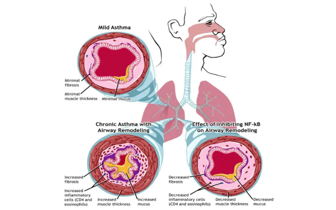
Eosinophilic Asthma
Eosinophilic asthma is a severe subtype of asthma, driven by high eosinophil levels. It often resists standard treatments and may not be allergy-related.

Eosinophilic Asthma
Eosinophilic asthma is a severe subtype of asthma, driven by high eosinophil levels. It often resists standard treatments and may not be allergy-related.
Infectious Mononucleosis (Mono)
Infectious mononucleosis (Mono), the “kissing disease,” is a common viral illness (EBV). Symptoms include sore throat, fever, fatigue, and swollen glands. Usually resolves on its own.
Causes of Eosinophilia (High Eosinophils)
High eosinophil count in blood. May indicate allergies, infections, or other conditions. Symptoms vary. Diagnosis via blood test.
Chronic Myeloid Leukemia Treatment Strategies
Chronic Myeloid Leukemia Treatment: TKIs are the main therapy, targeting the BCR::ABL1 gene. Chemotherapy & stem cell transplant are also used.
Neutrophilia (High Neutrophils) & Absolute Neutrophilia
Discover causes & implications of high neutrophils, from general neutrophilia to absolute neutrophilia, a key blood cell elevation.
Eosinophilic Esophagitis (EoE)
EoE: Chronic esophageal inflammation. Dysphagia, food impaction. Diagnosis: endoscopy, biopsies. Treatment: diet, meds, dilation.
AL Amyloidosis (Primary Amyloidosis)
AL amyloidosis occurs when misfolded light-chain proteins deposit in organs. Prompt treatment improves outcomes.


