Key Takeaways
An antibody identification antigram is a chart that shows which red cell antigens are present (+) or absent (0) on each reagent cell in a panel, allowing laboratories to identify an unknown red cell antibody.
- Interpretation works by inclusion and exclusion: positive reactions point to the antigen the antibody targets, and negative reactions rule out other antigens.
- The rule of three is the standard for a confident identification: at least 3 antigen-positive cells must react and at least 3 antigen-negative cells must not react, giving roughly 95% statistical confidence [1].
- The dosage effect can hide antibodies in the Rh, or minor blood groups like Kidd, Duffy, and MNS systems, so homozygous (double-dose) cells are preferred for ruling antibodies in or out [1,4].
- A negative autocontrol is needed before calling an alloantibody. A positive autocontrol points toward autoantibodies or recent transfusion and usually requires further workup [1,2].
- Correctly identifying the antibody allows the lab to choose antigen-negative donor units, which prevents acute and delayed hemolytic transfusion reactions and protects future pregnancies from hemolytic disease of the fetus and newborn [7,8].
Introduction
Blood transfusion saves lives every day, but giving incompatible blood can trigger a severe, sometimes fatal, hemolytic reaction. To prevent this, transfusion laboratories use a step-by-step process called antibody identification, and the central tool in that process is the antibody identification antigram.
This article walks you through what an antigram is, the principle behind it, and how to read one step by step. By the end, you should be able to follow the logic from reaction pattern to antibody name and understand why each rule exists.
What Is an Antigram?
An antigram is a grid. Each row represents one reagent red blood cell from a commercial panel, and each column represents a known antigen. A "+" means the antigen is present on that cell, and a "0" means it is absent [1].
The lab incubates the patient's serum or plasma with each panel cell, usually by the indirect antiglobulin test (IAT), a method that detects IgG antibodies bound to red cells. While traditional tube testing relies on visually observing cells agglutinate (clump), most modern laboratories utilize automated gel microcolumn assays (CAT) or solid-phase red cell adherence (SPRCA) testing. In SPRCA, a positive reaction is indicated by a diffuse layer of cells adhering to the bottom of the microplate well, rather than a clump [9]. The resulting reaction pattern is then read against the antigram, and the antigen the antibody is targeting can be deduced.
Principle of Antibody Identification Antigram Interpretation
Antibodies, also called immunoglobulins, are proteins made by the immune system that bind to specific antigens. Red blood cells display many antigens on their surface, organized into more than 40 blood group systems [3]. When a person is exposed to foreign red cells through transfusion or pregnancy, they may produce antibodies against antigens they do not carry themselves. These are alloantibodies.
The antigram allows us to match the pattern of positive and negative reactions to a single antigen profile. If every cell that reacts shares one antigen, and every cell that does not react lacks it, that antigen is almost certainly the target [1].
Interpretation Method
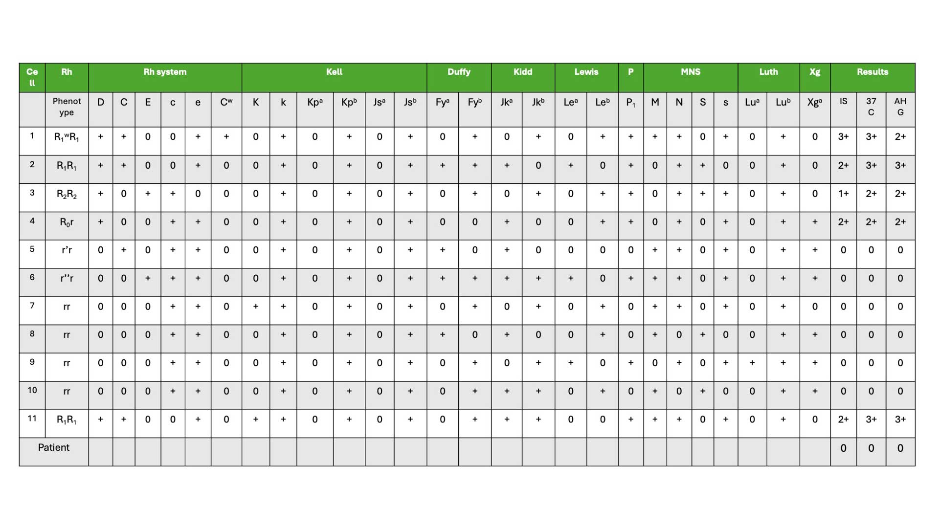
We will be using a worked example below to guide you through how to interpret the antibody identification antigram.
Step 1: Identify the Non-Reactive Cells
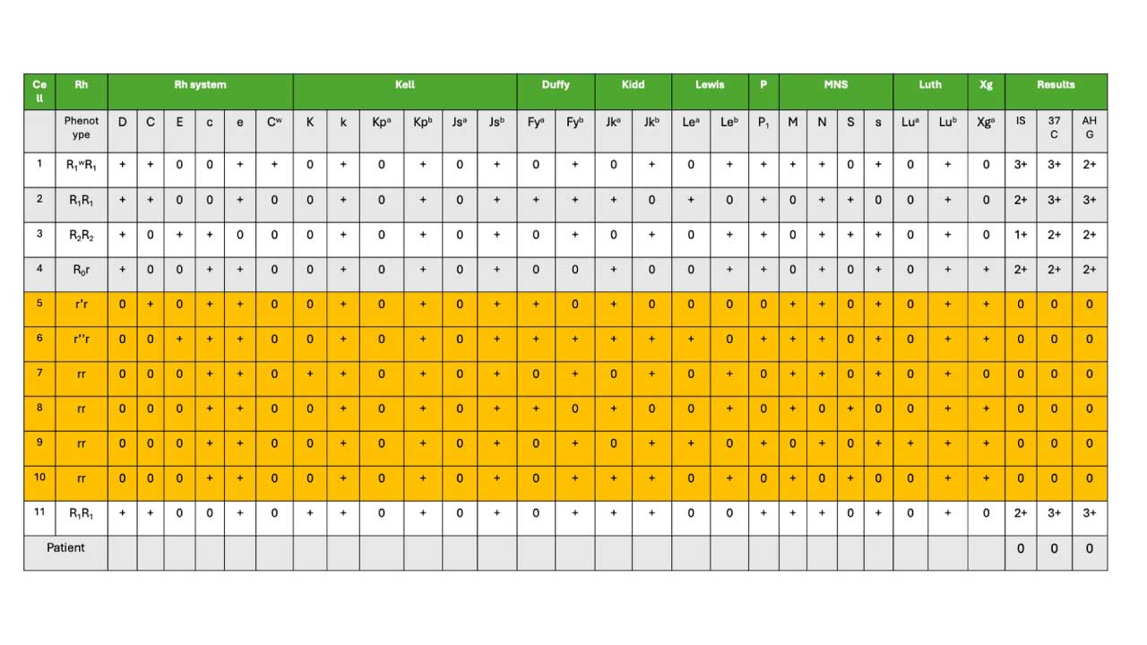
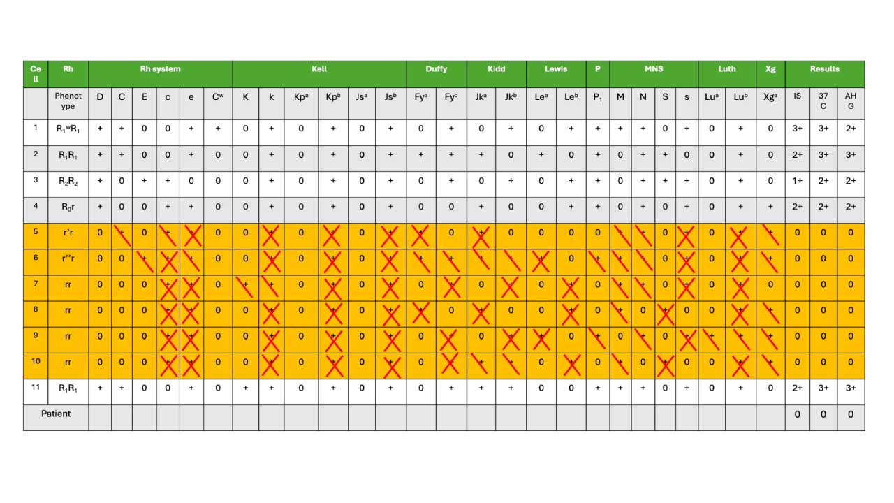
Start with the rows that show no reaction (0) across all test phases. In a typical 10-cell panel, several rows will be negative. In our worked example, cells 5–10 are non-reactive.
These cells are your starting point for exclusion. Whatever antigens they carry cannot be the antibody's target, because if the patient had that antibody, those cells would have reacted.
Step 2: Mark the Antigens to Exclude
For each non-reactive cell, look at every antigen marked "+" on that row and cross it off the suspect list. The convention is:
- Mark with "X" if the antigen is homozygous (present in double dose) on that cell.
- Mark with "\" if it is heterozygous (single dose).
Homozygous exclusions are stronger because of the dosage effect (more on this below).
Step 3: Apply the Rule of Three for Exclusion
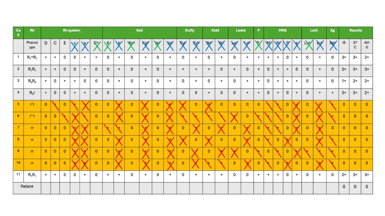
An antigen is confidently excluded once it has been crossed off three or more times, and ideally at least once on a homozygous (X) cell [1,2]. This is the rule of three, a statistical standard that gives approximately 95% confidence (p ≤ 0.05) that the antibody is not directed against that antigen.
Low-frequency or rare antigens (Cw, Kpa, Jsa, Lua, for example) can be crossed off without meeting the rule of three because they appear so rarely on donor cells that they are unlikely to cause clinical problems [1].
Step 4: Identify the Likely Antibody
After exclusions, look at the antigens still in play. Now check the positive reactions: do all the reactive cells share one remaining antigen? If they do, that is your suspect antibody.
In our worked example, the pattern points to anti-D. Every reactive cell carries the D antigen, and every non-reactive cell lacks it. It is critical to note that if anti-D is identified in a pregnant or recently postpartum patient, the laboratory and clinical team must investigate the patient's medical history to determine if this is an immune alloantibody or passive immunity resulting from a recent prophylactic administration of Rh immune globulin (e.g., RhIG or RhoGAM) [10].
Step 5: Confirm with the Rule of Three for Inclusion
To confirm that the antibody identified is anti-D, the following criteria must be fulfilled:
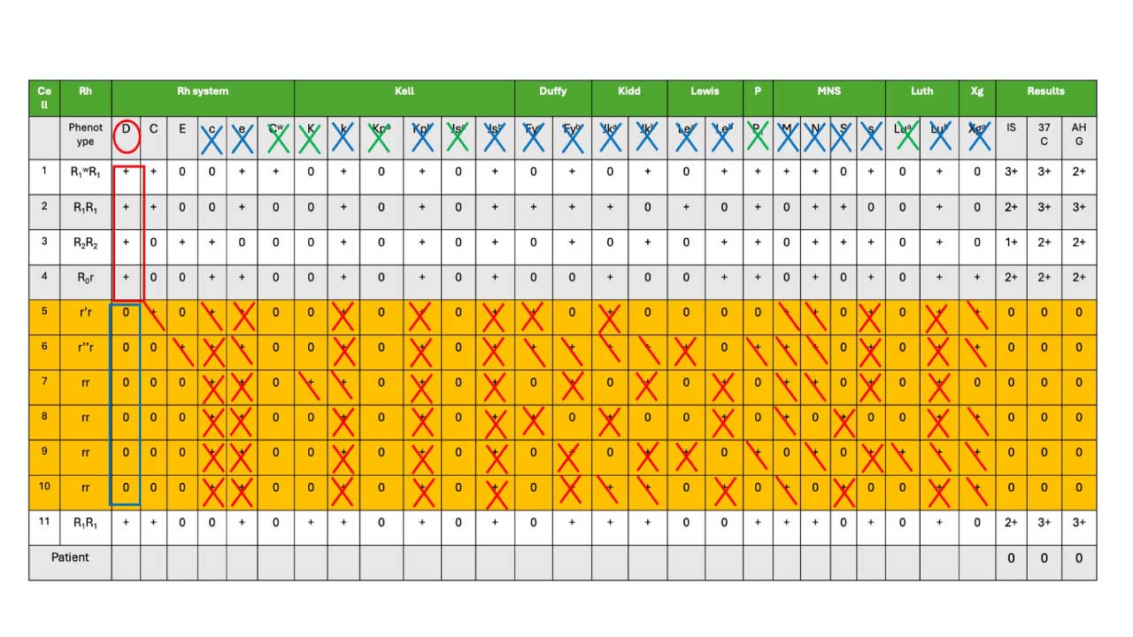
- Positive Reactions (Red Box): We need at least three red blood cells (RBCs) with the suspected antigen to be present and show a positive reaction (+) with the recipient's serum/plasma (marked in the red box).
- Negative Reactions (Blue Box): We also need at least three RBCs lacking the suspected antigen to exhibit a negative reaction (0) with the recipient's serum/plasma (marked in the blue box in cells 5-10).
- A negative autocontrol, confirming the reactions are not caused by autoantibodies.
If all three criteria are met, the rule of three is satisfied for inclusion as well as exclusion, and the antibody can be reported [1,2]. While the rule of three remains the historical AABB standard for 95% statistical confidence, some updated international guidelines and institutional protocols now accept a "rule of two" (two antigen-positive cells and two antigen-negative cells) for specific, frequently encountered single antibodies (such as anti-D) or when utilizing highly specific selected-cell panels to conserve limited reagent resources [11].
Clinically Significant Antibodies
It's very important to exclude common clinically significant antibodies like RhD, Duffy, MNSs, P, Kell, Kidd, and Lewis system antibodies through additional testing before concluding the antibody identification.
Step 6: Rule Out Other Clinically Significant Antibodies
Before finalizing the result, the lab must confirm there is no second antibody hiding underneath. Common clinically significant antibodies that should be excluded include those in the ABO, Rh, Kell, Kidd, Duffy, and Ss systems [1,2,7]. Lewis system antibodies are generally not considered clinically significant in adults and are usually noted but not pursued for transfusion compatibility [1,4].
If the panel cannot exclude a second antibody, additional testing with selected cells or enzyme-treated cells (ficin or papain) is used. Enzymes destroy some antigens (M, N, S, s, Fya, Fyb) while enhancing others (Rh, Kidd, Lewis, P1), which helps separate mixed antibody pictures. Furthermore, if antibodies to the Kell system are suspected in a complex mixture, laboratories may treat reagent cells with thiol-reducing agents like dithiothreitol (DTT) or AET. These chemicals destroy Kell system antigens without affecting other enzyme-sensitive antigens, allowing the lab to safely rule underlying antibodies in or out [1].
The Worked Example Antigram Result
The presence of the anti-D antibody (anti-D positive) is strongly suggested based on two key observations:
- Negative Autocontrol (AC): The autocontrol test, which uses the recipient's own red blood cells, is negative. This indicates the absence of self-directed antibodies (autoantibodies) that could potentially complicate the interpretation of the results.
- Anti-D Explains Reactions: The reactivity pattern observed in the antibody identification panel can be fully explained by the presence of anti-D. This means all the positive reactions in the panel are consistent with the expected behavior of anti-D binding to specific antigens on the test red blood cells.
Additional Considerations
- While anti-D is the most likely culprit, the possibility of other antibodies, particularly those targeting low-frequency antigens, cannot be entirely ruled out at this stage.
- Further testing may be necessary to definitively identify any additional antibodies, especially if the transfusion history suggests potential exposure to other blood group antigens.
- Ensure consistency between the antibody screen and identification results. This is a very important quality control step to verify the antibody identification reflects the actual antibodies present in the recipient's sample and minimizes the risk of transfusing incompatible blood due to a sample mix-up.
The Dosage Effect
Some antibodies react strongly only when the antigen is present in double dose. A heterozygous cell may give a weak or even negative reaction with the same antibody. This is the dosage effect, and it is classic in the Rh, Kidd, Duffy, and MNS blood group systems [1,4].
A practical example: imagine an anti-Jka antibody.
- Person 1's cells are Jk(a+b+) (heterozygous, single dose of Jka).
- Person 2's cells are Jk(a+b−) (homozygous, double dose of Jka).
- The antibody may react clearly with Person 2's cells but only weakly, or not at all, with Person 1's cells.
This is why labs prefer homozygous cells when ruling antibodies in or out. A negative result on a homozygous antigen-positive cell is much more reassuring than a negative on a heterozygous one.
When the Autocontrol Is Positive
A negative autocontrol points to an alloantibody. A positive autocontrol complicates the picture and suggests one of the following:
- A warm autoantibody.
- Recent transfusion with circulating donor cells.
- A drug-induced antibody.
- A positive direct antiglobulin test from another cause.
When this happens, the workup expands to include adsorption studies, elution, or a direct antiglobulin test (DAT) to separate auto from allo reactivity [1,2].
When the Antigram Does Not Give a Clear Answer
Real-world panels do not always produce a clean single-antibody pattern. Common challenges include:
- Multiple antibodies producing overlapping reactions. Selected-cell panels and enzyme-treated cells help untangle them [1].
- Antibodies to low-frequency antigens not represented on the standard panel.
- High-titer, low-avidity (HTLA) antibodies that react weakly across most cells.
- Patients with a positive DAT from autoimmune hemolytic anemia or recent transfusion.
In these situations, the case is often referred to a reference laboratory to resolve the discrepancy. Furthermore, modern clinical practice increasingly utilizes molecular red cell genotyping proactively rather than just reactively to solve complex antigrams. For chronically transfused populations, such as patients with sickle cell disease or thalassemia, baseline genotyping allows for prophylactic antigen matching to prevent alloimmunization before it begins [11].
Why This Matters Clinically
The whole point of antibody identification is patient safety. A missed or misidentified antibody can cause an acute or delayed hemolytic transfusion reaction, and in pregnant patients it can cause hemolytic disease of the fetus and newborn (HDFN) [7,8]. Anti-D and anti-K are the antibodies most commonly implicated in severe HDFN. Antibodies in the Kidd system are notorious for causing delayed reactions because their titers can fall below detection between transfusion episodes and then rise sharply on re-exposure [7].
Quality Checks Before Reporting
Before signing out an antibody identification result, the lab should confirm:
- The antibody screen and the panel results are consistent. A mismatch may indicate a sample mix-up.
- All clinically significant antibodies have been excluded.
- The rule of three is satisfied for both inclusion and exclusion.
- The autocontrol result has been interpreted.
- Any rare or low-frequency antigens have been considered.
Frequently Asked Questions (FAQs)
What is an antibody identification antigram used for?
An antibody identification antigram is a chart used in blood transfusion laboratories to figure out which red cell antibody a patient has produced. Each row represents a reagent red cell with a known antigen profile, and each column lists an antigen. By matching the patient's reaction pattern against the chart, the laboratory can identify the antibody and select compatible donor blood.
What does the rule of three mean in antibody identification?
The rule of three is a safety check used when interpreting an antigram. To call an antibody identified, the panel must show at least 3 antigen-positive cells that react with the patient's serum and at least 3 antigen-negative cells that do not react. This pattern gives about 95% statistical confidence that the antibody has been correctly identified.
What is the dosage effect and which blood groups show it?
The dosage effect is when an antibody reacts more strongly with red cells carrying two copies of an antigen (homozygous, double dose) than with red cells carrying one copy (heterozygous, single dose). It is most often seen with antibodies in the Rh, Kidd, Duffy, and MNS blood group systems. Recognizing dosage prevents false-negative interpretations.
What does it mean if the autocontrol is positive?
A positive autocontrol means the patient's serum is reacting with the patient's own red cells. Common causes include warm autoantibodies, a recent transfusion (with passively acquired antibody or drug effect), or a positive direct antiglobulin test. A positive autocontrol complicates interpretation and usually requires additional testing such as adsorption studies.
Which red cell antibodies are considered clinically significant?
Clinically significant antibodies are those that can cause hemolytic transfusion reactions or hemolytic disease of the fetus and newborn. Per AABB and BSH guidance, these include most antibodies in the ABO, Rh (D, C, c, E, e), Kell, Kidd (Jka, Jkb), Duffy (Fya, Fyb), and Ss systems. Lewis antibodies are generally not considered clinically significant in adults.
Why are homozygous cells preferred when ruling out antibodies?
Because of the dosage effect, an antibody might react with homozygous cells but not with heterozygous cells for the same antigen. Using a homozygous antigen-negative cell to rule out an antibody gives a more reliable negative result. If only heterozygous negative cells are available, additional testing may be needed.
Glossary of Related Medical Terms
- Antigram — A grid-style chart that lists which red cell antigens are present (+) or absent (0) on each reagent red cell in an antibody panel.
- Antibody panel — A set of about 10–20 reagent red cells with known antigen profiles, used to identify the specificity of an unknown antibody.
- Antigen — A molecule on the cell surface that can be recognized by an antibody. On red cells, antigens define the blood groups.
- Antibody (immunoglobulin) — A protein made by B cells that binds to a specific antigen.
- Agglutination — Visible clumping of red cells when antibody bridges them. A positive reaction in the panel.
- Autocontrol (AC) — A test that mixes the patient's serum with the patient's own red cells. A negative result rules out autoantibodies.
- Homozygous expression — The red cell carries two copies of a gene producing the antigen (double dose).
- Heterozygous expression — The red cell carries one copy (single dose). May give weaker reactions.
- Dosage effect — Stronger reaction with homozygous cells than heterozygous cells for the same antibody. Classic in Rh, Kidd, Duffy, and MNS systems.
- Rule of three — Statistical rule requiring at least 3 antigen-positive cells reacting and 3 antigen-negative cells not reacting, giving roughly 95% confidence in the antibody identification.
- Clinically significant antibody — An antibody capable of causing a transfusion reaction or hemolytic disease of the fetus and newborn. Includes most antibodies in the ABO, Rh, Kell, Kidd, Duffy, and Ss systems.
- Indirect antiglobulin test (IAT) — The serologic method used in panels. Patient serum is incubated with reagent cells, then anti-human globulin is added to detect IgG bound to red cells.
Disclaimer: This protocol is for educational purposes only. Local laboratory standard operating procedures take precedence. It is not intended to be a substitute for informed professional medical advice, diagnosis, or treatment. Always consult a qualified healthcare professional for clinical decision-making. While the information presented here is derived from credible medical sources and is believed to be accurate and up-to-date, it is not guaranteed to be complete or error-free. See additional information.
References
- Cohn, C. S., Delaney, M., Johnson, S. T., & Katz, L. M. (Eds.). (2023). Technical manual (21st ed.). AABB Press.
- British Committee for Standards in Haematology, Milkins, C., Berryman, J., Cantwell, C., Elliott, C., Haggas, R., Jones, J., Rowley, M., Williams, M., & Win, N. (2013). Guidelines for pre-transfusion compatibility procedures in blood transfusion laboratories. British Committee for Standards in Haematology. Transfusion medicine (Oxford, England), 23(1), 3–35. https://doi.org/10.1111/j.1365-3148.2012.01199.x
- Daniels, G. (2013). Human blood groups (3rd ed.). Wiley-Blackwell.
- Reid, M. E., Lomas-Francis, C., & Olsson, M. L. (2012). The blood group antigen factsbook (3rd ed.). Academic Press.
- Dean L. Blood Groups and Red Cell Antigens [Internet]. Bethesda (MD): National Center for Biotechnology Information (US); 2005. Available from: https://www.ncbi.nlm.nih.gov/books/NBK2261/
- Storry, J. R., Castilho, L., Chen, Q., Daniels, G., Denomme, G., Flegel, W. A., Gassner, C., de Haas, M., Hyland, C., Keller, M., Lomas-Francis, C., Moulds, J. M., Nogues, N., Olsson, M. L., Peyrard, T., van der Schoot, C. E., Tani, Y., Thornton, N., Wagner, F., Wendel, S., … Yahalom, V. (2016). International society of blood transfusion working party on red cell immunogenetics and terminology: report of the Seoul and London meetings. ISBT science series, 11(2), 118–122. https://doi.org/10.1111/voxs.12280
- Tormey, C. A., & Hendrickson, J. E. (2019). Transfusion-related red blood cell alloantibodies: induction and consequences. Blood, 133(17), 1821–1830. https://doi.org/10.1182/blood-2018-08-833962
- Delaney, M., Wendel, S., Bercovitz, R. S., Cid, J., Cohn, C., Dunbar, N. M., Apelseth, T. O., Popovsky, M., Stanworth, S. J., Tinmouth, A., Van De Watering, L., Waters, J. H., Yazer, M., Ziman, A., & Biomedical Excellence for Safer Transfusion (BEST) Collaborative (2016). Transfusion reactions: prevention, diagnosis, and treatment. Lancet (London, England), 388(10061), 2825–2836. https://doi.org/10.1016/S0140-6736(15)01313-6
- Harmening, D. M. (2018). Modern Blood Banking & Transfusion Practices (7th ed.). F.A. Davis Company. (Note: Chapter 9 thoroughly covers the transition from tube testing to Gel and SPRCA technologies).
- Practice Bulletin No. 181: Prevention of Rh D Alloimmunization. (2017). Obstetrics and gynecology, 130(2), e57–e70. https://doi.org/10.1097/AOG.0000000000002232
- Fasano, R. M., & Chou, S. T. (2016). Red Blood Cell Antigen Genotyping for Sickle Cell Disease, Thalassemia, and Other Transfusion Complications. Transfusion medicine reviews, 30(4), 197–201. https://doi.org/10.1016/j.tmrv.2016.05.011