Procedure-at-a-Glance
| Step | Action |
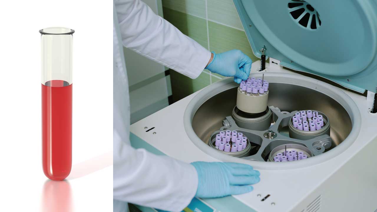
| 1. Collection | Collect 3mL EDTA blood for hemolysate preparation |
| 2. Separation | Centrifuge at 3000 rpm for 5 mins and discard the plasma later |
| 3. Washing | Wash RBCs 3x with saline |
| 4. Lysis | Add Lysis Agent |
| 5. Extraction | Vortex |
| 6. Purification | Centrifuge at 3000 rpm for 15 mins and filter |
| 7. Storage | Store or Freeze |
Introduction
Hemolysate, a suspension of lysed red blood cells, plays a crucial role in both Hb electrophoresis and alkaline denaturation tests. These tests rely on the biochemical properties of hemoglobin to differentiate between different types of hemoglobin and detect the presence of fetal blood.
Clinical Significance: Why Hemolysate Preparation is Essential
Preparing a pure hemolysate is the foundational step for diagnosing Hemoglobinopathies and Thalassemias. By lysing the red blood cells and removing the “debris” (stroma and plasma proteins), clinicians can isolate hemoglobin to identify specific genetic variants.
Screening for Hemoglobin Variants (HbS, HbC, HbE)
Hemoglobin electrophoresis is the gold standard for identifying structural abnormalities in the globin chains. The preparation of a clean hemolysate allows for the clear separation of:
- Hemoglobin S (HbS): The primary variant in Sickle Cell Disease. Identification is critical for early intervention in newborns to prevent splenic sequestration and vaso-occlusive crises.
- Hemoglobin C (HbC): Common in individuals of West African descent; it can cause mild hemolytic anemia or, when inherited with HbS (HbSC disease), a significant sickling disorder.
- Hemoglobin E (HbE): Prevalent in Southeast Asia; while often asymptomatic in trait form, it can cause severe anemia when inherited alongside Beta-thalassemia.
Diagnosis of Thalassemia
Thalassemias are characterized by the reduced production of normal hemoglobin chains. Hemolysate testing is used to quantify:
- Hemoglobin A2 (HbA2): Elevated levels are a hallmark indicator of Beta-Thalassemia Trait.
- Hemoglobin F (HbF): While normal in neonates, persistently high levels in adults can indicate Delta-Beta Thalassemia or Hereditary Persistence of Fetal Hemoglobin (HPFH).
Alkaline Denaturation & Hemolytic Disease of the Newborn (HDN)
The Alkaline Denaturation Test (or the Singher/Betke method) specifically exploits the fact that Fetal Hemoglobin (HbF) is highly resistant to denaturation in an alkaline environment, whereas adult Hemoglobin A is not.
- Evaluating Fetal-Maternal Hemorrhage: This test is vital in assessing “leaks” of fetal blood into maternal circulation.
- Hemolytic Disease of the Newborn: In cases of unexplained neonatal anemia or jaundice, quantifying HbF via hemolysate analysis helps clinicians differentiate between normal developmental patterns and pathological hemolysis.
Monitoring Treatment
For patients with Sickle Cell Disease who are treated with Hydroxyurea, preparing a hemolysate for regular electrophoresis or HPLC (High-Performance Liquid Chromatography) is necessary to monitor the therapeutic rise in HbF, which helps prevent red cell sickling.
Principle of Hemolysate Preparation
Preparation of hemolysate hinges on the controlled lysis of red blood cells (RBCs) to release their internal contents, particularly the protein hemoglobin without damaging the hemoglobin structure. This process, while seemingly simple, demands precise execution to ensure reliable results.
By introducing a detergent or hypotonic solution, the RBC membranes are gently broken down. These agents disrupt the phospholipid bilayer, the protective barrier surrounding the RBCs, causing them to burst and release their internal contents. The resulting solution, a mixture of hemoglobin and other cellular components, constitutes the raw hemolysate.
In some cases, further purification may be necessary. Filtration or gel filtration can be employed to remove cell debris and other unwanted components, resulting in a clear and concentrated hemolysate ideal for specific analyses.
Finally, the concentration of hemoglobin in the hemolysate must be carefully measured and adjusted to fall within the recommended range for each downstream test. Too little hemoglobin can lead to weak or invisible bands in electrophoresis, while too much can distort the results.
Materials
- EDTA peripheral blood sample
- Saline
- Distilled water
- Filter paper
- Glass tubes 5 ml
- Pipettes and micropipettes
- Vortex mixer
- Detergent or hypotonic solution (e.g., Triton X-100, ammonium chloride)
- Fine Pasteur pipette
- Optional: Toluene, carbon tetrachloride (CCl4), or chloroform
Protocol
- Collect 3 mL of fresh blood in an EDTA-anticoagulated tube.
- Transfer the blood to a glass tube.
- Centrifuge the blood at 3000 rpm (1006 g) for 5 minutes.
- Carefully remove the plasma layer without disturbing the red blood cells.
- Wash the red blood cells three times with saline solution.
- After each wash, centrifuge the sample according to step 3 and remove the supernatant.
- Following the final wash, note the volume of packed red blood cells.
- Choose the appropriate lysis method based on the desired application:
- Detergent Lysis: Add a volume of detergent solution (e.g., Triton X-100) according to the manufacturer’s instructions.
- Hypotonic Lysis: Add 0.75 times the volume of packed red blood cells with distilled water.
- Mix the lysed red blood cells with a vortex mixer for 2-3 minutes.
- (Optional) Add 0.5 times the volume of packed red blood cells with toluene, carbon tetrachloride (CCl4), or chloroform.
- Mix the solution vigorously with a vortex mixer for 2-3 minutes.
- Centrifuge the solution at 3000 rpm (1006 g) for 15 minutes.
- Using a fine Pasteur pipette, carefully remove the clear hemolysate layer located below the red cell membrane plug (stroma) or at the top of the solution (if using solvents).
- Wet a filter paper with distilled water to prevent complete absorption of the hemolysate.
- Filter the hemolysate to obtain a clear solution.
- Store the hemolysate at 4°C for immediate use.
- For long-term storage, aliquot the hemolysate and freeze at -80°C. Thaw frozen hemolysate on ice before use.
Interpretation
The clear and transparent hemolysate indicates successful lysis of the red blood cells and minimal contamination with other cellular components. The hemoglobin concentration is within the expected range for the patient’s age, sex, and other relevant clinical factors.
These results suggest that the hemolysate is suitable for use in further analyses, such as Hb electrophoresis and alkaline denaturation test. However, additional quality control procedures may be necessary depending on the specific downstream application.
Further Considerations:
- The measured hemoglobin concentration should be interpreted in the context of the patient’s clinical history and laboratory findings.
- Any deviations from the expected range should be investigated further to determine the underlying cause.
- The specific interpretation of the results may vary depending on the intended use of the hemolysate.
Note: This is a general example of a results and interpretation section for hemolysate preparation. The specific content and interpretation will vary depending on the individual patient and the specific testing procedures used.
There are several methods to measure the concentration of hemoglobin in the hemolysate, each with its own advantages and disadvantages:
| Method | Description | Advantages | Disadvantages |
| Spectrophotometry | This is the most common and widely available method. It utilizes the principle that hemoglobin absorbs light at a specific wavelength (540 nm for oxyhemoglobin). By measuring the absorbance of the hemolysate at this wavelength and comparing it to a standard curve, the hemoglobin concentration can be determined. | Fast and easy to perform. Requires minimal equipment. Relatively inexpensive. | May be inaccurate for high or low hemoglobin concentrations. Susceptible to interference from other substances in the hemolysate. |
| Cyanmethemoglobin method | This method involves converting hemoglobin to cyanmethemoglobin, a stable derivative with a unique absorption spectrum. By measuring the absorbance of the cyanmethemoglobin solution at a specific wavelength (540 nm), the hemoglobin concentration can be calculated. | More accurate than spectrophotometry for high and low hemoglobin concentrations. Less susceptible to interference from other substances. | More time-consuming than spectrophotometry. Requires additional reagents and equipment. |
| Automated hematology analyzer | Modern hematology analyzers can measure hemoglobin concentration directly using various technologies, such as flow cytometry or Coulter principle. | Fast and accurate. Requires minimal hands-on time. Provides additional blood cell parameters. | Requires specialized equipment. More expensive than other methods. |
Choosing the right method
The best method for measuring hemoglobin concentration in the hemolysate depends on various factors, including:
- Desired accuracy: If high accuracy is required, the cyanmethemoglobin method or an automated hematology analyzer may be preferred.
- Available resources: If resources are limited, spectrophotometry may be a more practical option.
- Sample volume: Some methods require larger sample volumes than others.
- Downstream analysis: Certain methods may be preferred depending on the subsequent analysis performed on the hemolysate.
It is important to consult the relevant laboratory manual or reagent instructions for detailed procedures and specific considerations when choosing and using a method for measuring hemoglobin concentration.
Common Challenges in Hemolysate Preparation
Even a standardized protocol can yield poor results if variables aren’t tightly controlled. Below are the most common issues encountered during hemolysate preparation and how to resolve them.
| Technical Issue | Observation | Potential Cause | Corrective Action |
| Lipid Interference | Hemolysate is turbid, milky, or cloudy. | Incomplete removal of cell membranes (stroma) or high lipid content in patient blood. | Increase centrifugation time/speed OR perform a solvent extraction with toluene or chloroform. |
| Protein Contamination | “Smearing” or extra bands (like Albumin) on electrophoresis. | Inadequate washing of the red cell mass. | Ensure cells are washed 3 times with 0.85% saline. The supernatant of the 3rd wash must be colorless. |
| Sample Dilution | Final bands are too faint to quantify. | Too much distilled water added during the lysis step. | Maintain a strict ratio: 1 part packed RBCs to 0.75 parts distilled water. |
| Hemoglobin Oxidation | Sample turns a dark brownish-red (Methemoglobin). | Old blood sample (>1 week) or exposure to high temperatures during vortexing. | Use fresh EDTA samples. If centrifugation generates heat, use a refrigerated centrifuge set to 4°C. |
| Incomplete Lysis | Visible “clumps” or a red cell button remains after mixing. | Insufficient mechanical agitation or using saline instead of water for lysis. | Ensure you use distilled/deionized water (hypotonic). Vortex vigorously for 30–60 seconds. |
| Poor Separation | Hemolysate contains “floaters” or debris. | Fibrin clots in the original sample or poor filtration. | Always check the EDTA tube for micro-clots before starting. Filter the final hemolysate through Whatman No. 1 filter paper. |
Frequently Asked Questions (FAQs)
Can I use heparinized blood instead of EDTA for hemolysate preparation?
While EDTA is preferred for hemoglobin studies, heparin can be used; however, avoid using citrate as it may dilute the sample significantly and interfere with some electrophoresis buffers.
Why is it critical to wash the red cells three times with saline?
Washing removes plasma proteins (like albumin and globulins). If these proteins remain, they can create “background noise” or extra bands on an electrophoresis strip, leading to a misinterpretation of results.
What is the purpose of adding toluene or chloroform during the protocol?
These organic solvents help to further precipitate the red cell stroma (membranes) and lipids, resulting in a much clearer, “cleaner” hemolysate which is ideal for high-resolution testing.
How do I know if my hemolysate is too concentrated?
If the hemolysate is too dark/opaque, it may cause “streaking” on the electrophoresis gel. Most protocols recommend adjusting the final hemoglobin concentration to approximately 10 g/dL before application.
Can I use frozen whole blood to prepare a hemolysate?
No. Freezing whole blood causes uncontrolled lysis and prevents the proper washing of red cells. You must wash the cells before the final lysis and freezing step.
Glossary of Related Medical Terms
- Alkaline Denaturation: A biochemical test (like the Singer or Betke test) used to quantify Hemoglobin F by measuring its resistance to denaturation at a high pH.
- EDTA (Ethylenediaminetetraacetic acid): A common anticoagulant used in hematology that prevents clotting by chelating calcium.
- Hb Electrophoresis: A laboratory technique used to separate different types of hemoglobin molecules based on their electrical charge.
- Hemolysate: The liquid product resulting from the rupture (lysis) of red blood cell membranes, containing released hemoglobin.
- Hypotonic Solution: A solution with a lower solute concentration than the inside of a cell, causing water to enter the cell and lead to osmotic lysis.
- Packed Red Blood Cells (pRBCs): Red blood cells that have been separated from the plasma through centrifugation.
- Stroma: The empty “ghost” membranes of red blood cells left behind after hemoglobin has been released.
- Supernatant: The clear liquid overlying material deposited by settling, precipitation, or centrifugation.
Disclaimer: This protocol is intended for informational purposes only and may need to be modified depending on the specific laboratory procedures and patient circumstances. Always consult with a qualified healthcare professional for guidance. See additional information.