Introduction
The Wells' score is a clinical prediction tool used to estimate how likely a patient is to have a deep vein thrombosis (DVT) before any blood test or imaging is ordered. It works by assigning points to specific findings from the patient's history and physical exam, such as recent surgery, leg swelling, or a previous clot. The resulting Wells' score sorts patients into a "DVT unlikely" or "DVT likely" category, which then determines the next diagnostic step. This matters because DVT symptoms overlap heavily with far more common conditions like muscle strain or cellulitis, and imaging every patient with a swollen leg would be slow, costly, and unnecessary.
The tool was developed by Dr. Philip Wells and colleagues and has since been validated in multiple large studies [1,2]. It is used daily by emergency physicians, internists, general practitioners, and nurse practitioners, and it forms the backbone of most DVT diagnostic algorithms taught in medical and nursing programs.
The Wells' Score Calculator
Wells' Criteria Calculator for DVT
Select all clinical features that apply to calculate the pre-test probability.
Each finding contributes to the running total. A negative point for an equally likely alternative diagnosis reflects the fact that the tool is meant to weigh probability, not simply count risk factors.
Interpreting the Wells' Score
Clinicians typically use one of two scoring models.
Two-level model (most common in practice):
- Score ≤ 1 → DVT unlikely (about 5–10% of these patients actually have a clot)
- Score ≥ 2 → DVT likely (about 25–40% actually have a clot)
Three-level model (used in some settings):
- 0 points or fewer → Low probability
- 1 to 2 points → Moderate probability
- 3 or more points → High probability
A low Wells' score is not a diagnosis on its own. It is a starting point that tells the clinician which test to order next.
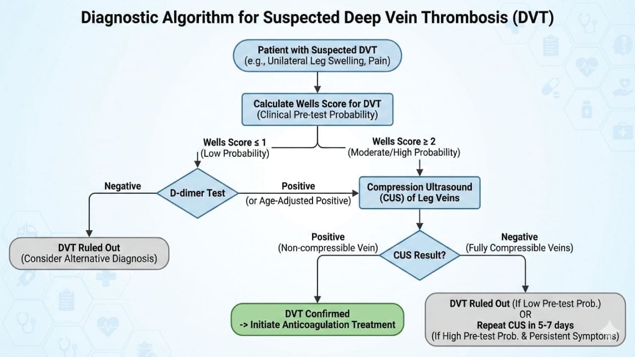
- If the score suggests DVT is unlikely, the next step is a D-dimer blood test. A normal D-dimer in this group is usually enough to rule out DVT without imaging [2]. For patients 50 years and older, modern protocols use an age-adjusted D-dimer cutoff (Patient's Age × 10 µg/L) to prevent false positives and safely avoid unnecessary ultrasounds in older adults [3].
- If the score suggests DVT is likely, D-dimer is skipped, and the patient goes straight to a compression ultrasound, since a normal D-dimer would not be reliable enough to rule out a clot in this higher-risk group.
Case Scenarios
Scenario 1: Low probability, ruled out with a blood test
A 34-year-old office worker presents with mild calf discomfort after a long car trip. She has no history of cancer, surgery, or immobilization, and no visible swelling on exam. Her only point comes from mild localized tenderness along her calf.
Interpretation: Wells' score = 1 → DVT unlikely. A D-dimer is ordered and comes back normal. No further testing is needed, and DVT is confidently ruled out.
Scenario 2: High probability, straight to ultrasound
A 68-year-old man is seen two weeks after hip replacement surgery. He reports a swollen, tender right calf. On exam, the entire leg is swollen, there is pitting edema, and tenderness follows the deep venous system.
Points: recent major surgery (+1), entire leg swollen (+1), pitting edema (+1), localized tenderness (+1)
Interpretation: Wells' score = 4 → DVT likely. He proceeds directly to compression ultrasound, which confirms a proximal DVT. Anticoagulation is started that day.
Scenario 3: Moderate probability, negative ultrasound needs a second look
A 55-year-old woman with active breast cancer presents with calf swelling and tenderness after a long flight.
Points: active cancer (+1), localized tenderness (+1), calf swelling ≥ 3 cm (+1)
Interpretation: Wells' score = 3 → DVT likely.
Her initial compression ultrasound is negative, but given her high pre-test probability, a single negative scan is not enough to fully exclude DVT. She is asked to return for a repeat ultrasound in 5–7 days to check whether a smaller, distal clot has propagated proximally, and no anticoagulation is started in the meantime unless symptoms worsen.
Limitations of the Wells' Score
The tool was validated specifically for the lower limbs; it does not apply to suspected upper-extremity DVT, which needs separate assessment. Furthermore, the Wells' score is not validated for pregnant patients. Because normal pregnancy inherently causes leg swelling and elevates D-dimer levels, using the Wells' score in this population is inaccurate; alternative clinical prediction tools like the LEFt rule must be used instead [4]. The Wells' score also relies partly on subjective clinical judgment, particularly the "alternative diagnosis at least as likely" item, which can vary between examiners. Because of this, the Wells' score works best as one part of a structured pathway alongside D-dimer testing and imaging, not as a standalone decision-maker.
Disclaimer: This article is intended for educational and informational purposes only. It is not intended to be a substitute for informed professional medical advice, diagnosis, or treatment. While the information presented here is derived from credible medical sources and is believed to be accurate and up-to-date, it is not guaranteed to be complete or error-free. See additional information.
References
- Wells, P. S., Anderson, D. R., Rodger, M., Forgie, M., Kearon, C., Dreyer, J., Kovacs, G., Mitchell, M., Lewandowski, B., & Kovacs, M. J. (2003). Evaluation of D-dimer in the diagnosis of suspected deep-vein thrombosis. The New England journal of medicine, 349(13), 1227–1235. https://doi.org/10.1056/NEJMoa023153
- Kearon, C., de Wit, K., Parpia, S., et al. (2022). Diagnosis of deep vein thrombosis with D-dimer adjusted to clinical probability: Prospective diagnostic management study. BMJ, 376, e067378. https://doi.org/10.1136/bmj-2021-067378
- Le Gal, G., Robert-Ebadi, H., Thiruganasambandamoorthy, V., Moustafa, F., Penaloza, A., Catella, J., Grenot, M. C., Visser, S., Mazzolai, L., Plumacker, A., Barco, S., Lang, E., Tagalakis, V., Deroche, C., Garnett, M., Hulme, J., Roy, P. M., Ghuysen, A., Couturaud, F., Engelberger, R., … ADJUST-DVT Investigators (2026). Age-Adjusted D-Dimer Cutoff Levels to Rule Out Deep Vein Thrombosis. JAMA, 335(5), 416–424. https://doi.org/10.1001/jama.2025.21561
- Chan, W. S., Lee, A., Spencer, F. A., Crowther, M., Rodger, M., Ramsay, T., & Ginsberg, J. S. (2009). Predicting deep venous thrombosis in pregnancy: out in "LEFt" field?. Annals of internal medicine, 151(2), 85–92. https://doi.org/10.7326/0003-4819-151-2-200907210-00004