Procedure-at-a-Glance
The BCB (Brilliant Cresyl Blue) stain is a supravital dye, meaning it stains living, unfixed red blood cells. It binds residual ribosomal RNA in reticulocytes and precipitates unstable hemoglobins like HbH [1,2].
- Mix equal parts EDTA blood and filtered stain (BCB or NMB).
- Incubate 10–15 min (NMB) or 15–30 min (BCB) at room temperature.
- Gently mix the tube.
- Make a thin film on a clean glass slide.
- Air dry (or low-cool hair dryer).
- Examine slide under oil immersion, ×100
Why This Stain Still Matters
Walk into any hematology lab today and most reticulocyte counts are done on an automated analyzer in seconds. So why teach a manual stain that takes 30 minutes? Two reasons. First, the BCB stain remains the go-to method for screening Hemoglobin H disease which is a job automated counters do not do. Second, the manual NMB method is still the comparator that validates every automated analyzer in use [1].
This guide walks through the principle, the protocol, and the interpretation, with the modern parameters (IRF, RPI, ARC) you will see on real reports.
A Quick Refresher: Why Reticulocytes Have RNA
A reticulocyte is a red blood cell that has just lost its nucleus but still carries leftover ribosomes. For roughly one to two days after leaving the marrow, it keeps making small amounts of hemoglobin from this residual RNA [3]. That RNA is invisible on a routine fixed smear. Supravital dyes catch it before the cell finishes maturing, which is why we apply the dye to living blood, not a fixed slide.
Principle of Supravital Stain
Supravital staining uses basic (cationic) dyes that cross the membranes of living red cells and bind to acidic intracellular structures, mainly ribosomal RNA. The dye clumps the RNA into a visible blue mesh called the reticulum [1,2]. Because the cells are still alive when stained, the dye also nudges unstable hemoglobins toward precipitation which is how Heinz bodies and HbH inclusions become visible.
This is the key contrast with Wright-Giemsa: Wright-Giemsa fixes (kills) cells first with methanol, so RNA stays diffuse and the reticulum never forms. Supravital first, fix never.
How BCB Differs From NMB Chemically
Brilliant Cresyl Blue is an oxazine dye. It tends to vary between batches and acts as a stronger oxidant, which is exactly what makes it good at forcing unstable hemoglobins to precipitate.
New Methylene Blue is a thiazine dye. It is more chemically pure, more soluble, and stains the reticulum a darker, sharper blue. That sharpness is why two technicians counting the same NMB slide will usually agree, and why CLSI H44-A2 chose NMB as the comparator method for validating automated reticulocyte counters [1].
| Feature | Brilliant Cresyl Blue (BCB)Oxazine dye | New Methylene Blue (NMB)Thiazine dye |
|---|---|---|
| Chemistry & Reagent Quality | ||
| Chemical class | Oxazine; batch variability | Thiazine; highly purified |
| Reference status | Used for HbH screening | Comparator method in CLSI H44-A2 [1] |
| Crystal precipitation | Frequent; filter every use | Less frequent; still filter |
| Morphologic Appearance | ||
| Reticulum appearance | Mid-to-light blue, sometimes faint | Dark blue, high contrast |
| RBC background | Pale blue-green | Pale greenish-yellow |
| Clinical Utility | ||
| Main clinical strength | HbH inclusion detection (α-thalassemia) | Routine reticulocyte counting |
| Oxidant action | Stronger; precipitates unstable HbH | Milder; cleaner RNA staining |
| Heinz bodies | Excellent | Excellent |
| Quality & Reproducibility | ||
| Inter-observer agreement | Lower (lighter staining) | Higher |
Where BCB Fits in a Modern Lab
Most labs now run flow-cytometry-based reticulocyte counts using fluorescent dyes such as thiazole orange. These are faster, more precise, and provide the Immature Reticulocyte Fraction (IRF) automatically [4,5]. Manual BCB and NMB remain essential for three jobs: Hemoglobin H screening, method comparison and quality control for automated counters, and resource-limited or field laboratories without flow cytometry [1].
Method differs slightly according to the manufacturer's protocol.
Materials
- 1.0% BCB or 1.0% NMB (filtered immediately before use)
- 1 mL EDTA peripheral blood
- 1 small glass test tube (10 × 75 mm)
- Pasteur pipette
- Dry incubator at 37°C (for the HbH protocol)
- Hair dryer (cool setting)
- Clean glass slides
Protocol
Routine Reticulocyte Count
Use this protocol with either NMB (preferred) or BCB.
- Filter a small aliquot of the dye through filter paper. Dye crystals look like inclusions and will fool the counter.
- Place 2–3 drops of filtered stain in a small test tube. Add an equal volume (2–3 drops) of well-mixed EDTA blood.
- Mix gently by flicking the bottom of the tube.
- Incubate at room temperature. NMB: 10–15 minutes. BCB: 15–30 minutes.
- Resuspend the mixture, then place one drop on a clean slide and make a thin smear.
- Dry the slide quickly using a cool hair dryer or by air-drying tilted.
- View under oil immersion at ×100. Do not fix the slide first as fixation destroys the appearance.
After 15 minutes, both reticulum and Heinz bodies are visible. Heinz bodies stain dark blue-purple at the membrane edge.
Specialized Protocol: BCB for HbH Inclusions
This is the screening test for α-thalassemia (Hemoglobin H disease). The principle is mild oxidative stress: BCB pushes unstable β4 tetramers (HbH) to precipitate inside the cell [2,7].
- Mix BCB stain and EDTA blood at a 1:1 ratio in a test tube.
- Incubate at 37°C in a dry incubator.
- Prepare smears at multiple time points: 1, 2, and 4 hours. Some HbH inclusions precipitate quickly; others need longer oxidative exposure to appear, so multiple time points improve sensitivity [7].
- Air dry or use a cool hair dryer.
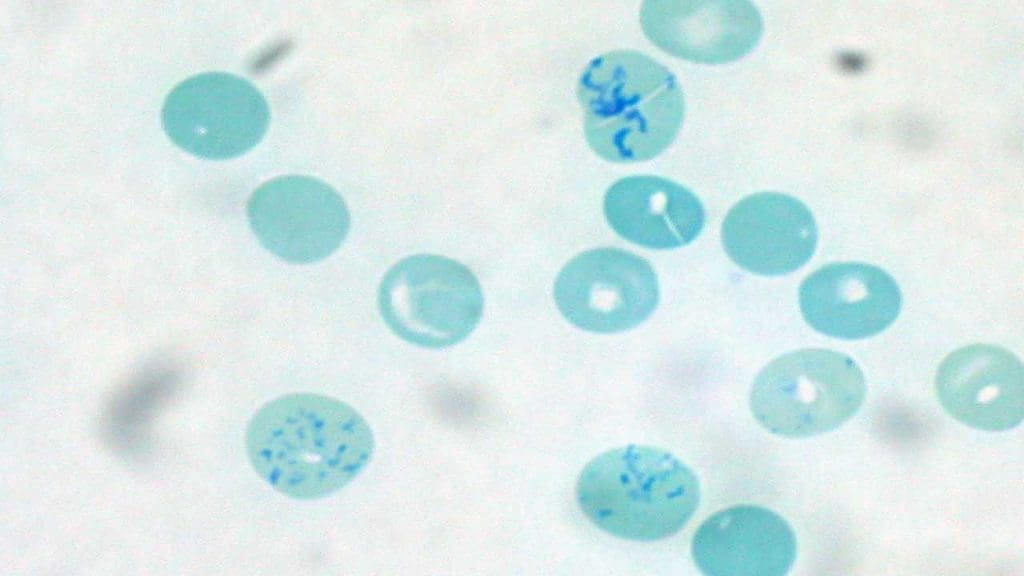
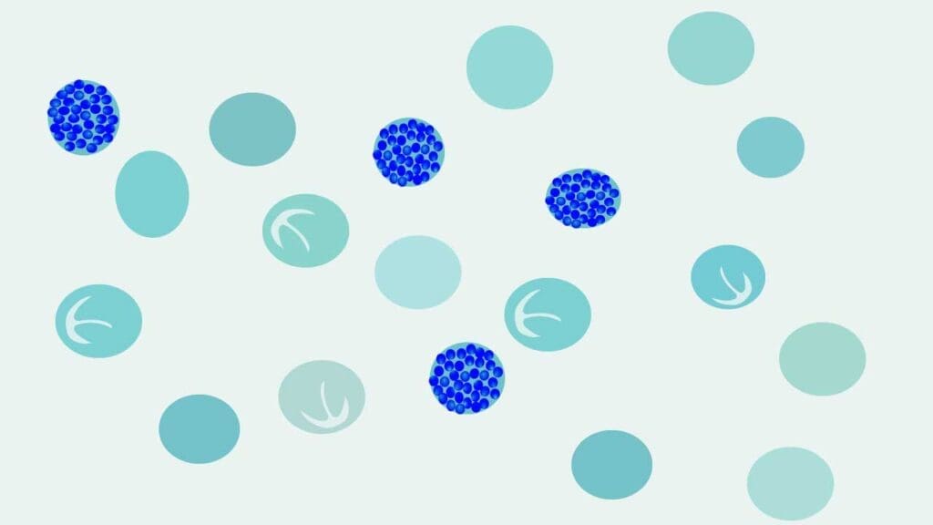
- Examine under oil immersion at ×100. Look for the "golf-ball" pattern: multiple small, evenly distributed greenish-blue dots throughout the red cell.
Preanalytical note
The HbH test must use fresh blood, ideally within 6–8 hours of collection. Stored EDTA samples lose reducing capacity, and HbH may precipitate passively in the tube before staining starts, distorting results.
Why HbH Looks Like a Golf Ball
In α-thalassemia, the patient cannot make enough α-globin chains. The β-globin chains they do make have no α-partners to pair with, so four β-chains stick together to form HbH (β4) [2]. β4 is unstable. Under the mild oxidative stress of BCB, it denatures and precipitates as many small clumps spread across the cell producing the "golf-ball" or "raspberry" appearance. NMB can also reveal HbH but less reliably, which is why BCB stays the screening dye of choice.
Interpretation
Normal Reference Ranges
- Adults: approximately 50–100 × 10⁹/L (0.5–2.5%) [3]
- Newborns and cord blood: 2–6%
The "Anemia Trap": Why Percentage Alone Misleads
Reticulocyte percentage is a ratio. In severe anemia, fewer mature red cells make the percentage look high even when marrow output has not changed. To get the true picture, use ARC and RPI.
Absolute Reticulocyte Count (ARC)
The ARC is the actual number of reticulocytes per volume of blood. It removes the percentage distortion entirely.
ARC = (Reticulocyte % / 100) × RBC count
Example: A patient with a 2.0% reticulocyte count and an RBC count of 4.0 × 10⁶/µL has an ARC of 0.02 × 4,000,000 = 80,000/µL.
Normal range: approximately 25–75 × 10⁹/L (20,000–100,000/µL) [3].
Reticulocyte Production Index (RPI)
The RPI is the gold standard for clinical decision-making in anemia. It corrects for two things: the patient's anemia, and shift reticulocytes — young cells released early from a stressed marrow that linger in circulation for 2–3 days instead of the usual 1 day. Without correction, you would count those cells multiple days in a row and overestimate marrow output.
RPI = Corrected Retic % ÷ Maturation Factor
Corrected Retic % = Observed Retic % × (Patient's Hct ÷ 45%)
| Patient Hematocrit | Maturation Factor (days) |
|---|---|
| 45% | 1.0 |
| 35% | 1.5 |
| 25% | 2.0 |
| 15% | 2.5 |
| Marrow Response | RPI | What It Means | Typical Conditions |
|---|---|---|---|
| Regenerative | > 3.0 | Marrow producing RBCs at high rate to compensate for loss | Hemolytic anemia (G6PD) Sickle cell AIHA Acute hemorrhage Recent iron or B12 therapy |
| Inadequate | 2.0–3.0 | Marrow trying but not keeping up | Mild blood loss Early marrow recovery |
| Non-regenerative | < 2.0 | Marrow damaged or starved of building blocks | Iron-deficiency anemia Aplastic anemia Megaloblastic anemia (B12/folate) MDS |
Immature Reticulocyte Fraction (IRF)
IRF is the proportion of the youngest, most RNA-rich reticulocytes in circulation, reported automatically by modern hematology analyzers using fluorescent staining [4,5]. It rises before the total reticulocyte count rises, which makes it the earliest blood signal of marrow recovery.
Where IRF earns its keep:
- Detecting marrow recovery after chemotherapy or stem cell transplant
- Monitoring response to iron, B12, or folate replacement
- Distinguishing aplastic from regenerative anemia early in workup
- Following erythropoietin response in chronic kidney disease
Reference intervals vary by analyzer, but IRF is typically 2–14% in healthy adults [5].
Reticulocyte Hemoglobin Content (CHr / RET-He)
CHr (also reported as RET-He) measures the actual hemoglobin inside reticulocytes — a real-time snapshot of how well iron is reaching the marrow [4]. It drops within days of iron deficiency, faster than ferritin or transferrin saturation reflect changes. It is now widely used to screen iron deficiency in children and to monitor iron status in dialysis patients.
Frequently Asked Questions (FAQs)
Why can't reticulocytes be counted on a routine Wright-Giemsa stain?
Wright-Giemsa stain fixes cells with methanol before staining, which stops the RNA inside reticulocytes from clumping. You may see a bluish-gray tint called polychromasia, but the diagnostic mesh-like reticulum will not appear. Only supravital stains such as BCB or NMB, applied to living cells, can precipitate the RNA into a visible network for counting.
When should a lab choose BCB over NMB?
Choose BCB when screening for Hemoglobin H disease (a form of α-thalassemia). BCB is a stronger oxidant and reliably precipitates unstable HbH into the characteristic "golf-ball" pattern of multiple small blue dots inside red cells. For routine reticulocyte counting, NMB is preferred because its staining is darker, sharper, and more reproducible between observers.
What is the Immature Reticulocyte Fraction (IRF) and why does it matter?
IRF is the percentage of the youngest, most RNA-rich reticulocytes in circulation, reported by modern automated hematology analyzers. It rises before the total reticulocyte count rises, making it the earliest blood marker of bone marrow recovery for example, after chemotherapy, stem cell transplant, or iron and B12 replacement. A rising IRF signals that the marrow is responding to treatment.
Why is the reticulocyte percentage misleading in anemia?
The percentage compares reticulocytes to mature red cells. In severe anemia, there are fewer mature cells, so the percentage looks falsely high even if marrow output is unchanged. Two corrections fix this: the Absolute Reticulocyte Count (ARC), which gives the true number per microliter, and the Reticulocyte Production Index (RPI), which also corrects for early-released "shift" reticulocytes that linger in the blood for 2–3 days instead of 1.
Can BCB or NMB stain Howell-Jolly bodies or Pappenheimer bodies?
They may show faintly, but these are not what supravital stains are designed for. Howell-Jolly bodies are DNA remnants and stain best on a routine Wright-Giemsa smear. Pappenheimer bodies are iron granules and need a Perls (Prussian blue) iron stain for confirmation. Supravital stains target RNA (reticulocytes) and denatured hemoglobin (Heinz bodies, HbH inclusions).
Why does the HbH inclusion test require fresh blood?
HbH precipitation depends on the metabolic state of living red cells reacting with the dye's mild oxidative push. Once EDTA blood is stored, cells become depleted of glutathione and other reducing systems. Some HbH precipitates passively in the tube before staining begins, while other unstable hemoglobin populations no longer respond. Best practice is to perform HbH screening within 6–8 hours of collection.
Glossary of Related Medical Terms
- Supravital stain: A dye applied to fresh, living cells (not fixed with alcohol) so it can interact with structures inside metabolically active cells.
- Reticulocyte: A young red blood cell, just released from the bone marrow, that still contains leftover ribosomal RNA and matures within about 1–2 days.
- Reticulum: The blue mesh of clumped RNA seen inside a reticulocyte after supravital staining.
- Erythropoiesis: The production of red blood cells in the bone marrow, regulated mainly by the hormone erythropoietin.
- Polychromasia: The bluish-gray tint of young red cells on a Wright-Giemsa stain.
- Heinz bodies: Clumps of denatured hemoglobin at the cell membrane, usually from oxidative stress (for example, in G6PD deficiency).
- HbH (Hemoglobin H): An abnormal hemoglobin made of four β-globin chains (β4), formed when α-globin is deficient (α-thalassemia).
- HbH inclusions: "Golf-ball" pattern of multiple small blue dots across the red cell after BCB staining.
- Absolute Reticulocyte Count (ARC): The actual number of reticulocytes per microliter of blood.
- Reticulocyte Production Index (RPI): A corrected reticulocyte value accounting for anemia and shift reticulocytes.
- Immature Reticulocyte Fraction (IRF): The youngest, most RNA-rich reticulocytes — an early marker of marrow recovery.
- Reticulocyte Hemoglobin Content (CHr / RET-He): Hemoglobin inside reticulocytes; a real-time marker of functional iron supply.
- Fixation: Chemical preservation (usually methanol) of cells. Supravital staining must happen before fixation.
Disclaimer: This protocol is for educational purposes only. Local laboratory standard operating procedures take precedence. It is not intended to be a substitute for informed professional medical advice, diagnosis, or treatment. Always consult a qualified healthcare professional for clinical decision-making. While the information presented here is derived from credible medical sources and is believed to be accurate and up-to-date, it is not guaranteed to be complete or error-free. See additional information.
References
- Clinical and Laboratory Standards Institute. (2004). Methods for reticulocyte counting (automated blood cell counters, flow cytometry, and supravital dyes); approved guideline (2nd ed., CLSI document H44-A2). CLSI.
- Bain, B. J. (2022). Blood cells: A practical guide (6th ed.). Wiley-Blackwell.
- Bain, B. J., Bates, I., & Laffan, M. A. (2016). Dacie and Lewis practical haematology (12th ed.). Elsevier.
- Piva, E., Brugnara, C., Spolaore, F., & Plebani, M. (2015). Clinical utility of reticulocyte parameters. Clinics in laboratory medicine, 35(1), 133–163. https://doi.org/10.1016/j.cll.2014.10.004
- Buttarello M. (2016). Laboratory diagnosis of anemia: are the old and new red cell parameters useful in classification and treatment, how?. International journal of laboratory hematology, 38 Suppl 1, 123–132. https://doi.org/10.1111/ijlh.12500
- Carr, J. H. (2021). Clinical hematology atlas (6th ed.). Elsevier.
- Pan, L. L., Eng, H. L., Kuo, C. Y., Chen, W. J., & Huang, H. Y. (2005). Usefulness of brilliant cresyl blue staining as an auxiliary method of screening for alpha-thalassemia. The Journal of laboratory and clinical medicine, 145(2), 94–97. https://doi.org/10.1016/j.lab.2004.11.019